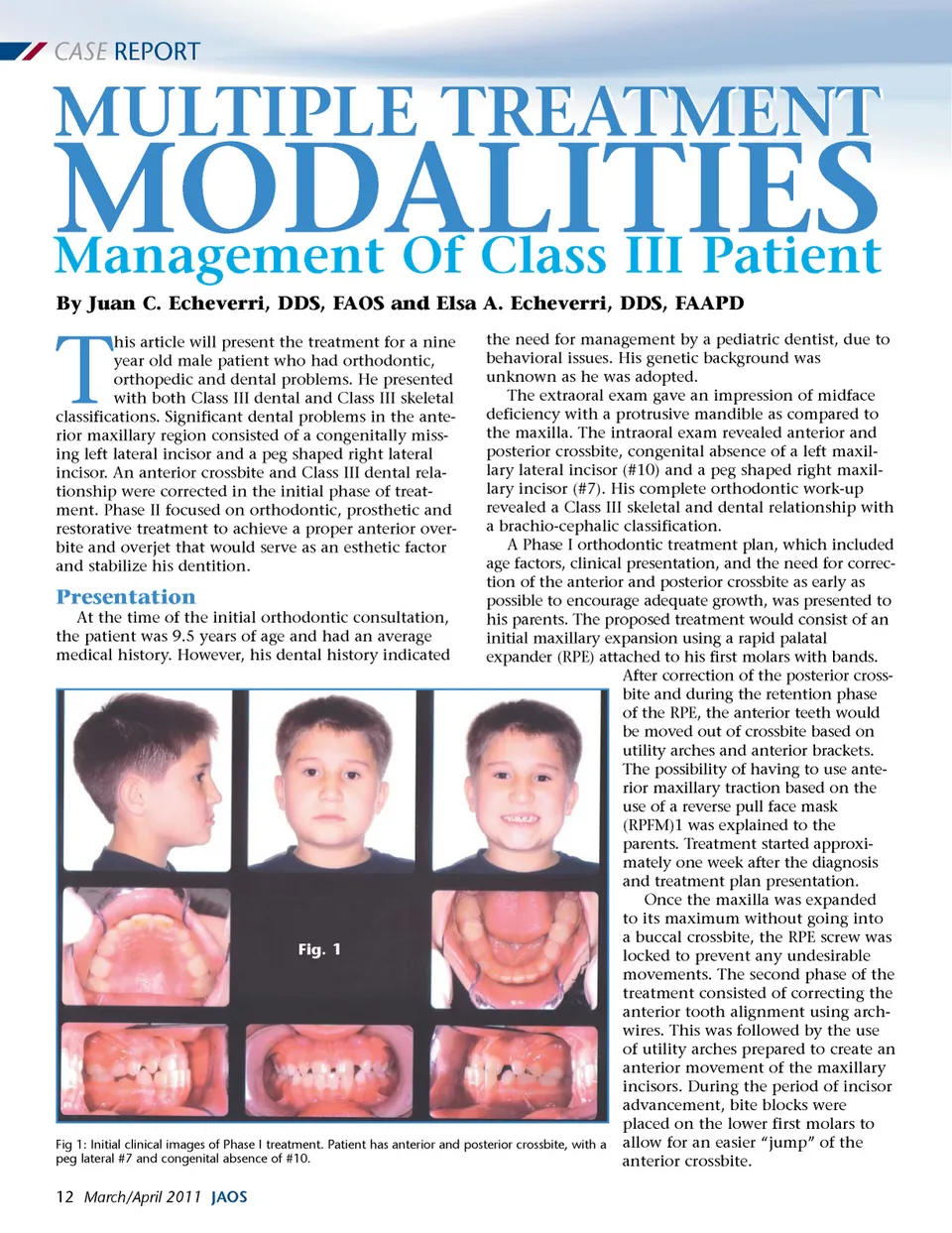
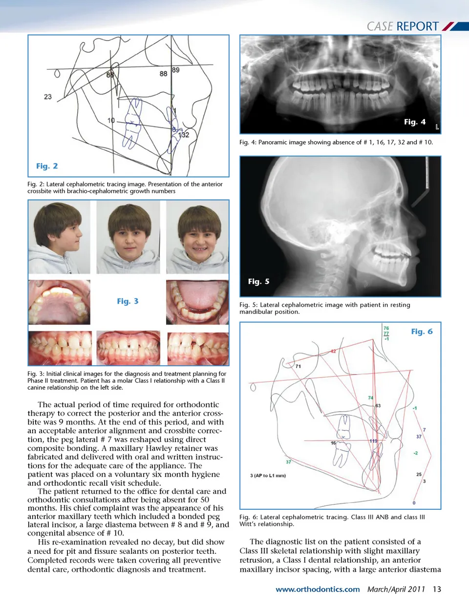
CASE REPORT Fig. 4 Fig. 4: Panoramic image showing absence of # 1, 16, 17, 32 and # 10. Fig. 2 Fig. 2: Lateral cephalometric tracing image. Presentation of the anterior crossbite with brachio-cephalometric growth numbers Fig. 5 Fig. 3 Fig. 5: Lateral cephalometric image with patient in resting mandibular position. Fig. 6 Fig. 3: Initial clinical images for the diagnosis and treatment planning for Phase II treatment. Patient has a molar Class I relationship with a Class II canine relationship on the left side. The actual period of time required for orthodontic therapy to correct the posterior and the anterior cross-bite was 9 months. At the end of this period, and with an acceptable anterior alignment and crossbite correc-tion, the peg lateral # 7 was reshaped using direct composite bonding. A maxillary Hawley retainer was fabricated and delivered with oral and written instruc-tions for the adequate care of the appliance. The patient was placed on a voluntary six month hygiene and orthodontic recall visit schedule. The patient returned to the office for dental care and orthodontic consultations after being absent for 50 months. His chief complaint was the appearance of his anterior maxillary teeth which included a bonded peg lateral incisor, a large diastema between # 8 and # 9, and congenital absence of # 10. His re-examination revealed no decay, but did show a need for pit and fissure sealants on posterior teeth. Completed records were taken covering all preventive dental care, orthodontic diagnosis and treatment. Fig. 6: Lateral cephalometric tracing. Class III ANB and class III Witt’s relationship. The diagnostic list on the patient consisted of a Class III skeletal relationship with slight maxillary retrusion, a Class I dental relationship, an anterior maxillary incisor spacing, with a large anterior diastema www.orthodontics.com March/April 2011 13
Journal of the American Orthodontic Society March-April 2011: Page 13