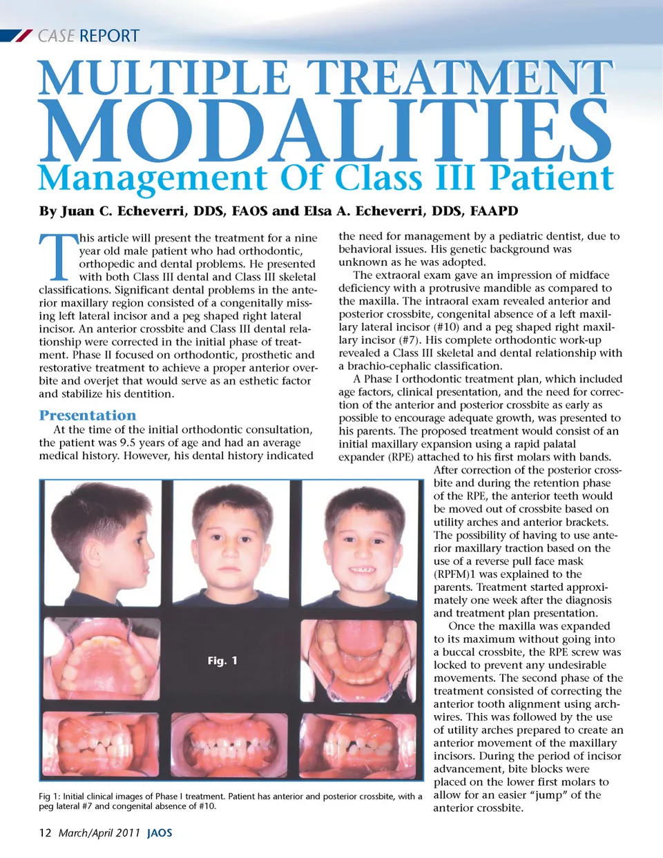
CASE REPORT MULTIPLE TREATMENT By Juan C. Echeverri, DDS, FAOS and Elsa A. Echeverri, DDS, FAAPD MODALITIES Management Of Class III Patient T the need for management by a pediatric dentist, due to behavioral issues. His genetic background was unknown as he was adopted. The extraoral exam gave an impression of midface deficiency with a protrusive mandible as compared to the maxilla. The intraoral exam revealed anterior and posterior crossbite, congenital absence of a left maxil-lary lateral incisor (#10) and a peg shaped right maxil-lary incisor (#7). His complete orthodontic work-up revealed a Class III skeletal and dental relationship with a brachio-cephalic classification. A Phase I orthodontic treatment plan, which included age factors, clinical presentation, and the need for correc-tion of the anterior and posterior crossbite as early as Presentation possible to encourage adequate growth, was presented to At the time of the initial orthodontic consultation, his parents. The proposed treatment would consist of an the patient was 9.5 years of age and had an average initial maxillary expansion using a rapid palatal medical history. However, his dental history indicated expander (RPE) attached to his first molars with bands. After correction of the posterior cross-bite and during the retention phase of the RPE, the anterior teeth would be moved out of crossbite based on utility arches and anterior brackets. The possibility of having to use ante-rior maxillary traction based on the use of a reverse pull face mask (RPFM)1 was explained to the parents. Treatment started approxi-mately one week after the diagnosis and treatment plan presentation. Once the maxilla was expanded to its maximum without going into a buccal crossbite, the RPE screw was Fig. 1 locked to prevent any undesirable movements. The second phase of the treatment consisted of correcting the anterior tooth alignment using arch-wires. This was followed by the use of utility arches prepared to create an anterior movement of the maxillary incisors. During the period of incisor advancement, bite blocks were placed on the lower first molars to Fig 1: Initial clinical images of Phase I treatment. Patient has anterior and posterior crossbite, with a allow for an easier “jump” of the peg lateral #7 and congenital absence of #10. anterior crossbite. his article will present the treatment for a nine year old male patient who had orthodontic, orthopedic and dental problems. He presented with both Class III dental and Class III skeletal classifications. Significant dental problems in the ante-rior maxillary region consisted of a congenitally miss-ing left lateral incisor and a peg shaped right lateral incisor. An anterior crossbite and Class III dental rela-tionship were corrected in the initial phase of treat-ment. Phase II focused on orthodontic, prosthetic and restorative treatment to achieve a proper anterior over-bite and overjet that would serve as an esthetic factor and stabilize his dentition. 12 March/April 2011 JAOS
Journal of the American Orthodontic Society March-April 2011: Page 12