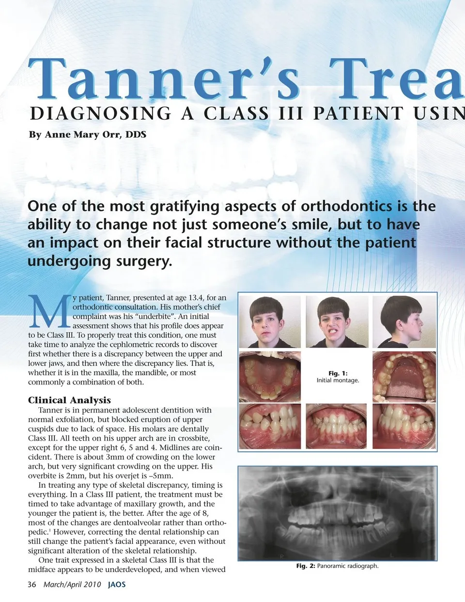
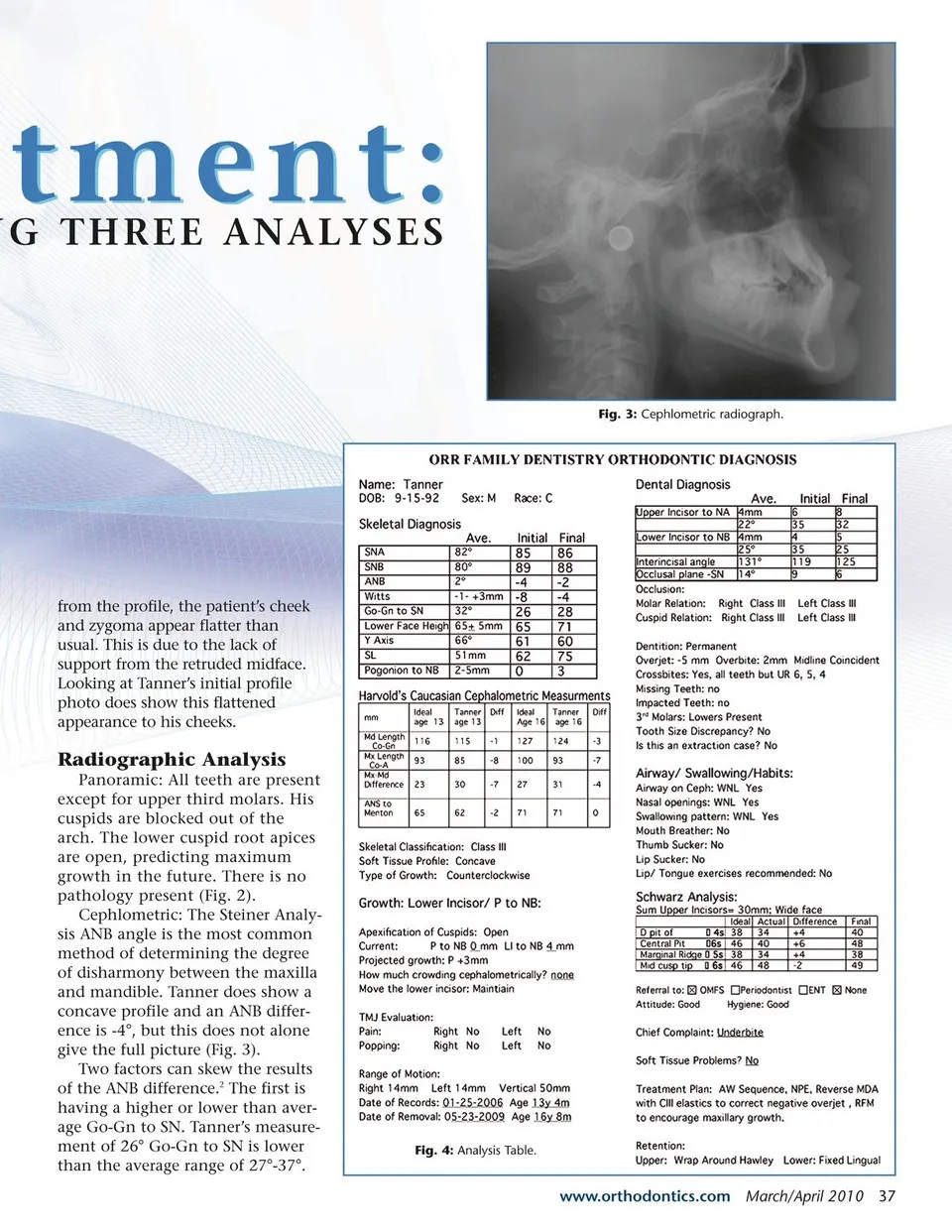
atment: NG THREE ANALYSES tment: Fig. 3: Cephlometric radiograph. from the profile, the patient’s cheek and zygoma appear flatter than usual. This is due to the lack of support from the retruded midface. Looking at Tanner’s initial profile photo does show this flattened appearance to his cheeks. Radiographic Analysis Panoramic: All teeth are present except for upper third molars. His cuspids are blocked out of the arch. The lower cuspid root apices are open, predicting maximum growth in the future. There is no pathology present (Fig. 2). Cephlometric: The Steiner Analy- sis ANB angle is the most common method of determining the degree of disharmony between the maxilla and mandible. Tanner does show a concave profile and an ANB differ- ence is -4°, but this does not alone give the full picture (Fig. 3). Two factors can skew the results of the ANB difference.2 The first is having a higher or lower than aver- age Go-Gn to SN. Tanner’s measure- ment of 26° Go-Gn to SN is lower than the average range of 27°-37°. Fig. 4: Analysis Table. www.orthodontics.com March/April 2010 37
Journal of the American Orthodontic Society March - April 2010: Page 37