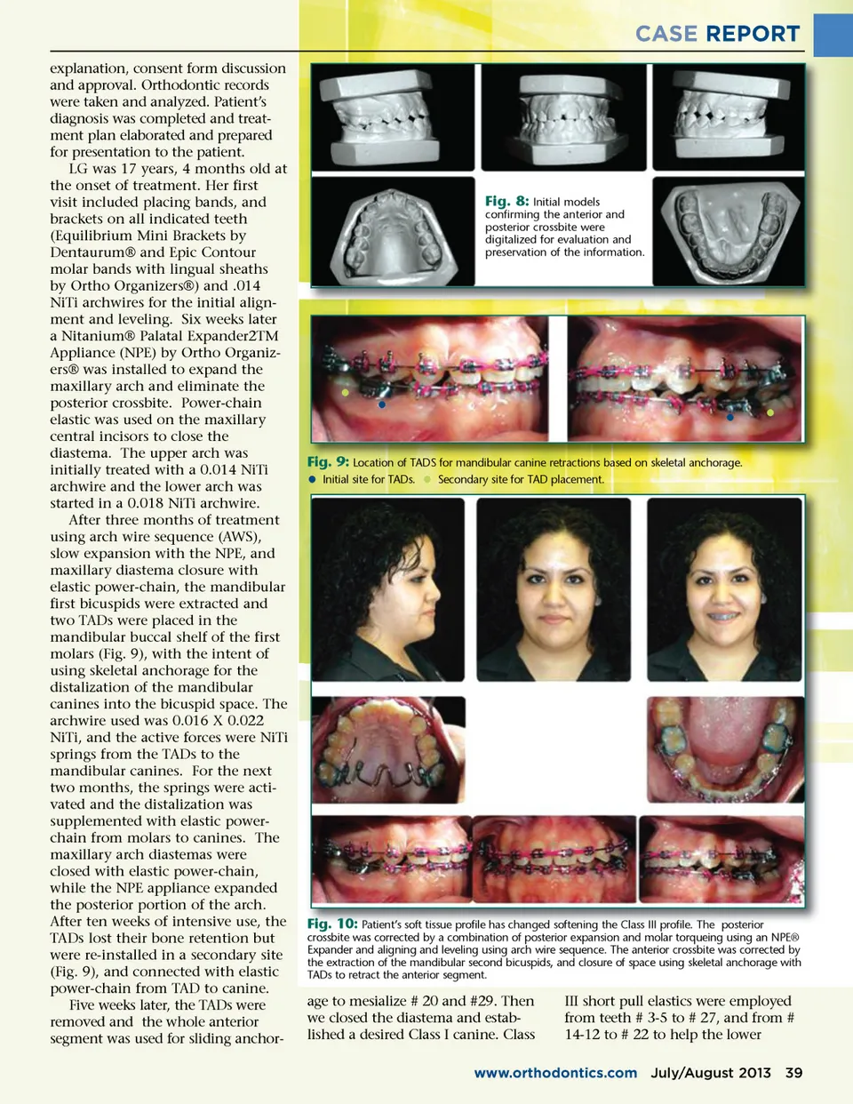
CASE REPORT explanation, consent form discussion and approval. Orthodontic records were taken and analyzed. Patient’s diagnosis was completed and treat-ment plan elaborated and prepared for presentation to the patient. LG was 17 years, 4 months old at the onset of treatment. Her first visit included placing bands, and brackets on all indicated teeth (Equilibrium Mini Brackets by Dentaurum® and Epic Contour molar bands with lingual sheaths by Ortho Organizers®) and .014 NiTi archwires for the initial align-ment and leveling. Six weeks later a Nitanium® Palatal Expander2TM Appliance (NPE) by Ortho Organiz-ers® was installed to expand the maxillary arch and eliminate the posterior crossbite. Power-chain elastic was used on the maxillary central incisors to close the diastema. The upper arch was initially treated with a 0.014 NiTi archwire and the lower arch was started in a 0.018 NiTi archwire. After three months of treatment using arch wire sequence (AWS), slow expansion with the NPE, and maxillary diastema closure with elastic power-chain, the mandibular first bicuspids were extracted and two TADs were placed in the mandibular buccal shelf of the first molars (Fig. 9), with the intent of using skeletal anchorage for the distalization of the mandibular canines into the bicuspid space. The archwire used was 0.016 X 0.022 NiTi, and the active forces were NiTi springs from the TADs to the mandibular canines. For the next two months, the springs were acti-vated and the distalization was supplemented with elastic power-chain from molars to canines. The maxillary arch diastemas were closed with elastic power-chain, while the NPE appliance expanded the posterior portion of the arch. After ten weeks of intensive use, the TADs lost their bone retention but were re-installed in a secondary site (Fig. 9), and connected with elastic power-chain from TAD to canine. Five weeks later, the TADs were removed and the whole anterior segment was used for sliding anchor-Fig. 8: Initial models confirming the anterior and posterior crossbite were digitalized for evaluation and preservation of the information. • • • • Fig. 9: Location of TADS for mandibular canine retractions based on skeletal anchorage. • Initial site for TADs. • Secondary site for TAD placement. Fig. 10: Patient’s soft tissue profile has changed softening the Class III profile. The posterior crossbite was corrected by a combination of posterior expansion and molar torqueing using an NPE® Expander and aligning and leveling using arch wire sequence. The anterior crossbite was corrected by the extraction of the mandibular second bicuspids, and closure of space using skeletal anchorage with TADs to retract the anterior segment. age to mesialize # 20 and #29. Then we closed the diastema and estab-lished a desired Class I canine. Class III short pull elastics were employed from teeth # 3-5 to # 27, and from # 14-12 to # 22 to help the lower www.orthodontics.com July/August 2013 39
Journal of the American Orthodontic Society July-August 2013 Buyer's Guide: Page 39