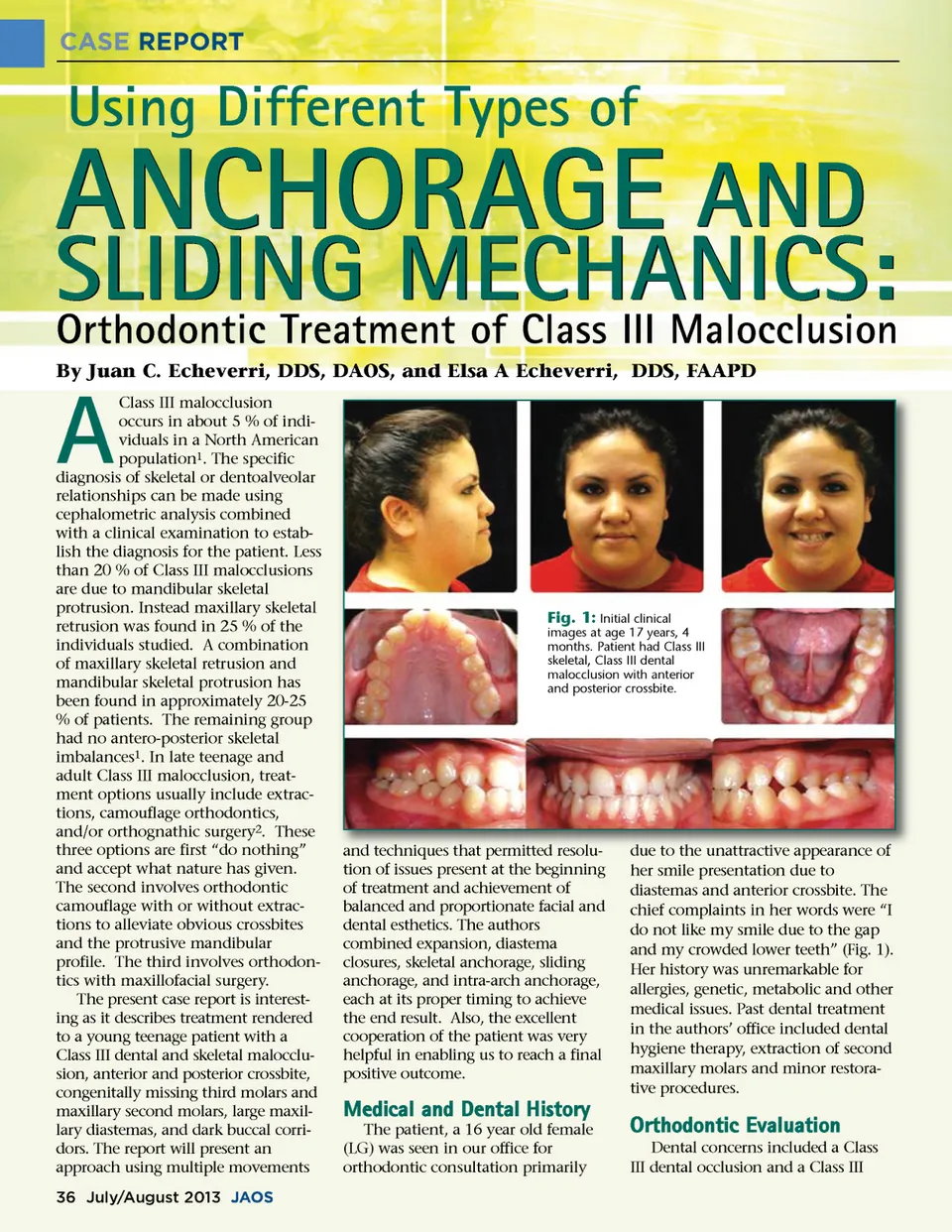
CASE REPORT Fig. 2: Maxillary second molars in ectopic position resembling wisdom teeth with no apparent opposing teeth due to Class III malocclusion. Image taken prior to the orthodontic consultation. Wisdom teeth were congenitally missing. Fig. 4: Initial cephalometric image. Observe the difference in proportion between the maxilla and the mandible. Upper lip symmetries with face and lower lip are slightly protrusive due to position of the mandibular incisors. Radiographic Analysis The panoramic image showed all permanent teeth present except for congenitally missing third molars and maxillary second molars (Fig. 3). A cephalometric radiograph was taken (Fig. 4) and traced using Gerety trac-ing analysis (Fig. 5) and confirmed with a Ricketts tracing analysis (Fig. 6). The tracing revealed a Class III skeletal classification based on 0 ANB and -8 Wits Appraisal. The mandibu-lar plane angle of 35˚, the Y-axis of 66˚ and the S-L of 53.4 mm indicated she had grown with a neutral clockwise growth tendency. Her soft tissue profile showed upper lips in a good position, but lower lips slightly protru-sive, due to the advanced position of her mandibular incisors which were slightly beyond the soft tissue line. The analysis indicated a Class III skele-Fig. 3: Panoramic image nine months after removal of maxillary second molars showing complete healing of extraction sites. skeletal type that included an anterior and posterior crossbite, and dark buccal corridors. Clinical and radio-graphical examination revealed no dental decay. There were no temporo-mandibular joint problems, but obvi-ous skeletal and dental conditions in need of orthodontic correction. The clinical oral exam revealed misaligned maxillary incisors, a maxillary midline shifted toward the right, an anterior crossbite of the lateral incisors, and a bilateral posterior crossbite. The mandible had moderate crowding, more evident in the incisor and bicus-pid regions. The frontal smile revealed dark buccal corridors and a canted smile with a resulting higher deviated left side. The lack of maxil-lary second molars was not noticeable due to the fullness of the upper arch with the teeth present. There was no excessive anterior gingival exposure. At the time of her orthodontic records,( LG) had turned 17. (Fig. 1). prior history of headaches, joint dysfunction, pain, clenching or grinding. Clinical examination revealed no popping, clicking or crepitus. Joint screening was within normal limits, with a vertical open-ing of 50 mm, a left deviation of 9 mm, and right deviation of 8 mm. T MJ Examination The TMJ examination was within normal parameters with no Fig. 5: Gerety lateral cephalometric tracing. Note the good maxillary incisor position for proper lip support and the lower incisors in protrusive position creating an anterior crossbite, protrusive lower lip, and Class III dental position. The vertical jaw opening at the higher end of a mesocephalic classification, created a class I ANB skeletal classification. www.orthodontics.com July/August 2013 37
Journal of the American Orthodontic Society July-August 2013 Buyer's Guide: Page 37