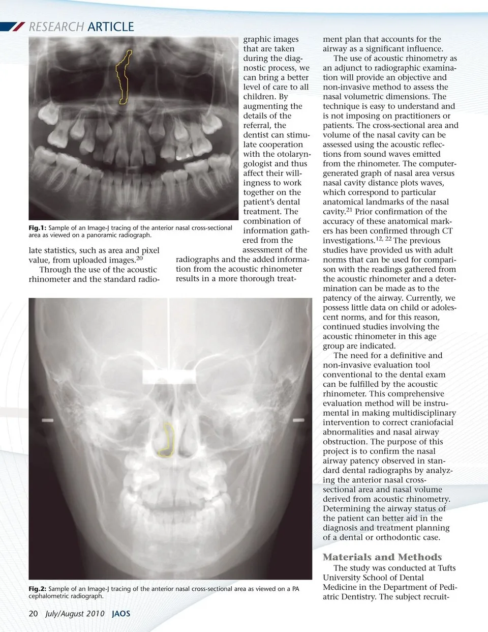
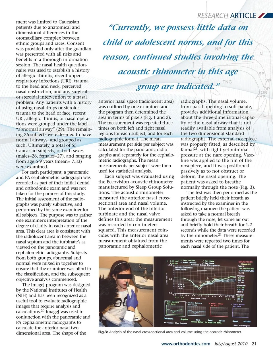
RESEARCH ARTICLE graphic images that are taken during the diag-nostic process, we can bring a better level of care to all children. By augmenting the details of the referral, the Fig.1: Sample of an Image-J tracing of the anterior nasal cross-sectional area as viewed on a panoramic radiograph. late statistics, such as area and pixel value, from uploaded images.20 Through the use of the acoustic rhinometer and the standard radio-dentist can stimu-late cooperation with the otolaryn-gologist and thus affect their will-ingness to work together on the patient’s dental treatment. The combination of information gath-ered from the assessment of the radiographs and the added informa-tion from the acoustic rhinometer results in a more thorough treat-ment plan that accounts for the airway as a significant influence. The use of acoustic rhinometry as an adjunct to radiographic examina-tion will provide an objective and non-invasive method to assess the nasal volumetric dimensions. The technique is easy to understand and is not imposing on practitioners or patients. The cross-sectional area and volume of the nasal cavity can be assessed using the acoustic reflec-tions from sound waves emitted from the rhinometer. The computer-generated graph of nasal area versus nasal cavity distance plots waves, which correspond to particular anatomical landmarks of the nasal cavity.21 Prior confirmation of the accuracy of these anatomical mark-ers has been confirmed through CT investigations.12, 22 The previous studies have provided us with adult norms that can be used for compari-son with the readings gathered from the acoustic rhinometer and a deter-mination can be made as to the patency of the airway. Currently, we possess little data on child or adoles-cent norms, and for this reason, continued studies involving the acoustic rhinometer in this age group are indicated. The need for a definitive and non-invasive evaluation tool conventional to the dental exam can be fulfilled by the acoustic rhinometer. This comprehensive evaluation method will be instru-mental in making multidisciplinary intervention to correct craniofacial abnormalities and nasal airway obstruction. The purpose of this project is to confirm the nasal airway patency observed in stan-dard dental radiographs by analyz-ing the anterior nasal cross-sectional area and nasal volume derived from acoustic rhinometry. Determining the airway status of the patient can better aid in the diagnosis and treatment planning of a dental or orthodontic case. Fig.2: Sample of an Image-J tracing of the anterior nasal cross-sectional area as viewed on a PA cephalometric radiograph. 20 July/August 2010 JAOS Materials and Methods The study was conducted at Tufts University School of Dental Medicine in the Department of Pedi-atric Dentistry. The subject recruit-
Journal of the American Orthodontic Society July-August 2010/Buyer's Guide: Page 20