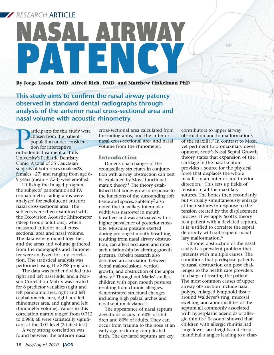
RESEARCH ARTICLE acteristic long and narrow face with midface deficiency.8 Chronic allergic rhinitis can cause structural changes at the cellular level that manifest as hyper-trophy of the lining of the nose. The normal ciliated columnar epithelium diminishes as mucus accumulates, clogging the epithe-lium, and damaging the cilia further with bacteria and viruses. The hypertrophic response of nasal tissue leads to reduced airway patency from obstructed nasal passages.9 The resultant altered breathing mode has been shown to change dental and facial “normal” growth.10,11 Another negative sequela to mouth breathing is an increase in the child’s cavity poten-tial now and throughout their life-time.12 Trask studied siblings who suffered from rhinitis and compared them to a set of siblings who did not have airway complications.13 He found the rhinitis sufferers had an increase in palatal height, a smaller angle between the mandibular plane and the mandibular incisors, increased over-jet, retrognathic mandibles, and increased lower facial height. In Linder-Aronson’s published report, he noted that mouth-breathers with prolonged airway obstruction exhibited cross-bites, narrow upper arches, long upper lip and shorter mandibular arch length.14 Malocclusions are either heredi-tary, functional, or a combination of both. Parents pass down certain skeletal and dental characteristics to their children through genetics. It is also well documented that abnormal functions, such as airway obstructions, thumb sucking habits, or chronic allergies can lead to mouth breathing. The open mouth postures adopted through this abnormal manner of breathing can alter the way in which teeth and faces grow. In patients who are still growing, obstruction of the upper airway can lead to excessive vertical facial growth, dental malformations, and aberrant oromaxillary development.15 Children between the ages of 6 and 9 years are particularly vulnera-ble to oromaxillary changes associ-ated with existing or evolving nasal airway complications. These patients are prime candidates for growth modification via intercep-tive orthodontic appliances. Further investigation is needed to deter-mine the impact of growth modifi-cation through orthodontics on nasal airway obstruction. Regard-less, a successful case must begin with accurate documentation of the dental and medical history. Resolu-tion and post-treatment retention can only be achieved if the clinician treats the primary etiology of the malocclusion as well as the airway obstruction. It is possible that chil-dren who undergo correction of malocclusions with appliances such as braces or palatal expansion, yet “The dentist can properly treat a child with nasal airway obstruction and subsequent dental malocclusions by working closely with the patient’s pediatrician and an otolaryngologist.” have continued airway obstruction, will have higher rates of dental relapse after their orthodontic treat-ment is complete.16 Therefore, in the process of diagnosing and correcting the dental malocclusion, the airway status is of keen interest to pediatric dentistry. A valid visualization and evalua-tion of the nasal airway, turbinates and nasal septum, can be performed through routine examination at the dental office via panoramic radio-graphs. Analysis of PA cephalometric radiographs, at times recorded at the onset of orthodontic record keeping, can enhance further diagnosis.17 According to a recent study by Corey, the acoustic rhinometer has shown reliability in noting changes in the patency of the nasal cavity in post-surgical cases.18 The rhinometer can be used, when available, to document nasal cavity dimensions pre-and post-orthodontic treatment in the developing child. The dentist can properly treat a child with nasal airway obstruction and subsequent dental malocclusions by working closely with the patient’s pediatrician and an otolaryngologist. A thorough medical questionnaire and clinical examination is impor-tant to understand the etiology of the problem. An accurate referral to the ear nose and throat specialist should include diagnostic informa-tion that is readily available to dentists from the standard initial radiographic exam. The cephalomet-ric and panoramic radiographs are very instrumental, but a more objec-tive standard is necessary to establish norms and baselines that limit human error from interpretation. Currently, dentists and orthodon-tists rely on subjective visual assess-ment of the airway extraorally and by analyzing panoramic radiographs and noting apparent structural and anatomical abnormalities. Once again, this current modality of analysis lacks a standardized norm and is subject to human error and thus imprecise. While some recent studies have attempted to bridge this knowledge gap, there is a need to investigate a comprehensive diagnostic tool that is non-invasive and can be coupled with standard dental imaging to formulate the best objective assessment possible. This is where we believe the use of the Acoustic Rhinometer by dentists and orthodontists can serve the greatest benefit to their patients. The acoustic rhinometer was developed in 1977 and has been used to measure nasal airflow and volume by the medical profession. Some of the earliest descriptions and applications of rhinometry where provided by Hilberg. Acoustic rhinometry is non-invasive and uses sound waves to calculate the nasal airway.19 ImageJ is a public domain Java image-processing program simi-lar to NIH Image. The program, released in 1997, is utilized to calcu-www.orthodontics.com July/August 2010 19
Journal of the American Orthodontic Society July-August 2010/Buyer's Guide: Page 19