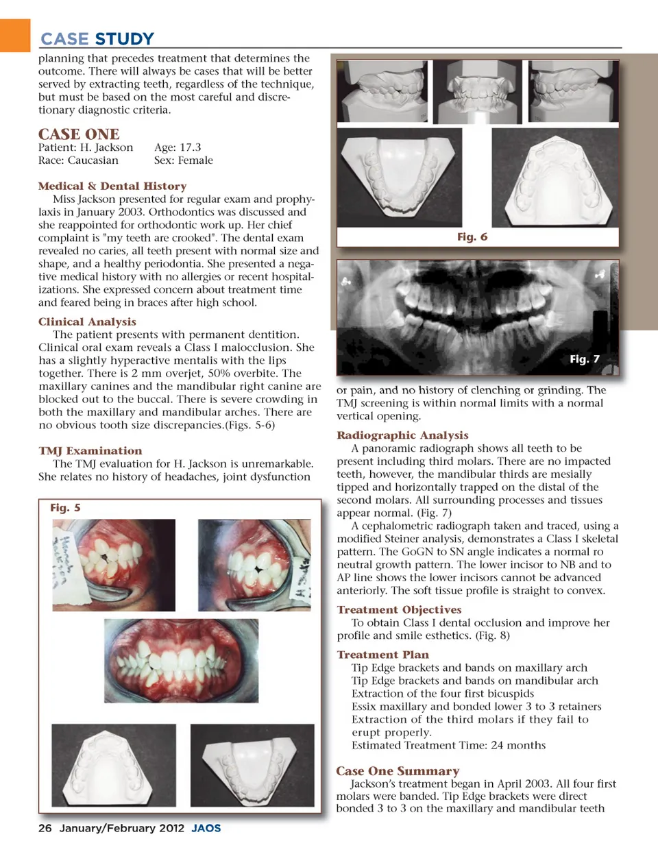
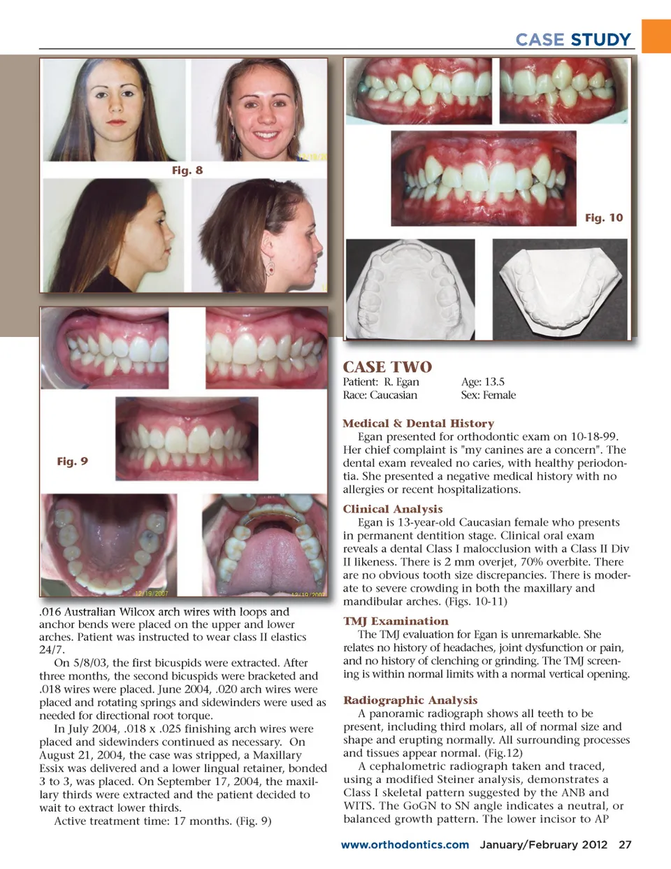
CASE STUDY Fig. 8 Fig. 10 CASE TWO Patient: R. Egan Race: Caucasian Age: 13.5 Sex: Female Fig. 9 Medical & Dental History Egan presented for orthodontic exam on 10-18-99. Her chief complaint is "my canines are a concern". The dental exam revealed no caries, with healthy periodon-tia. She presented a negative medical history with no allergies or recent hospitalizations. Clinical Analysis Egan is 13-year-old Caucasian female who presents in permanent dentition stage. Clinical oral exam reveals a dental Class I malocclusion with a Class II Div II likeness. There is 2 mm overjet, 70% overbite. There are no obvious tooth size discrepancies. There is moder-ate to severe crowding in both the maxillary and mandibular arches. (Figs. 10-11) TMJ Examination The TMJ evaluation for Egan is unremarkable. She relates no history of headaches, joint dysfunction or pain, and no history of clenching or grinding. The TMJ screen-ing is within normal limits with a normal vertical opening. Radiographic Analysis A panoramic radiograph shows all teeth to be present, including third molars, all of normal size and shape and erupting normally. All surrounding processes and tissues appear normal. (Fig.12) A cephalometric radiograph taken and traced, using a modified Steiner analysis, demonstrates a Class I skeletal pattern suggested by the ANB and WITS. The GoGN to SN angle indicates a neutral, or balanced growth pattern. The lower incisor to AP www.orthodontics.com January/February 2012 27 .016 Australian Wilcox arch wires with loops and anchor bends were placed on the upper and lower arches. Patient was instructed to wear class II elastics 24/7. On 5/8/03, the first bicuspids were extracted. After three months, the second bicuspids were bracketed and .018 wires were placed. June 2004, .020 arch wires were placed and rotating springs and sidewinders were used as needed for directional root torque. In July 2004, .018 x .025 finishing arch wires were placed and sidewinders continued as necessary. On August 21, 2004, the case was stripped, a Maxillary Essix was delivered and a lower lingual retainer, bonded 3 to 3, was placed. On September 17, 2004, the maxil-lary thirds were extracted and the patient decided to wait to extract lower thirds. Active treatment time: 17 months. (Fig. 9)
Journal of the American Orthodontic Society January-February 2012: Page 27