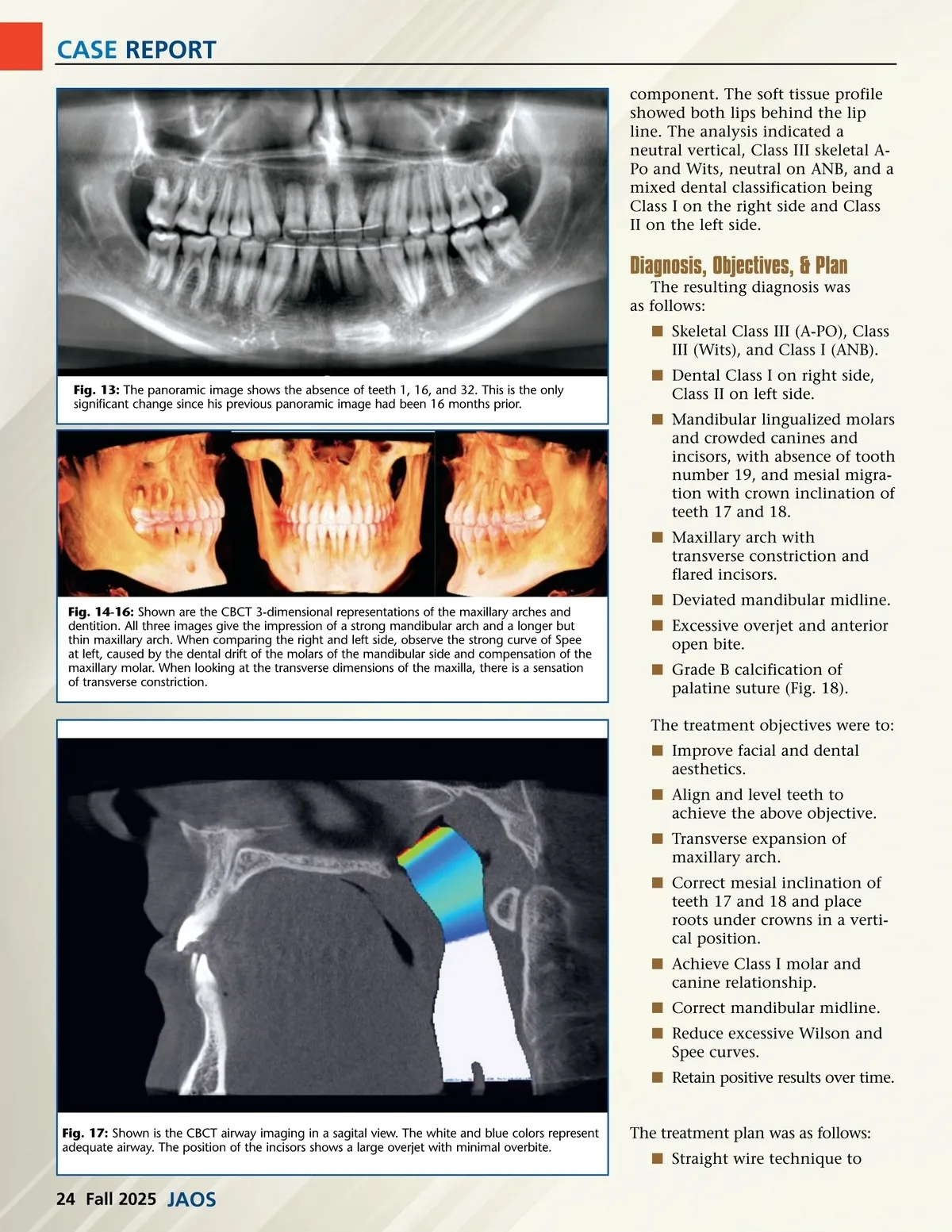
CASE REPORT component. The soft tissue profile showed both lips behind the lip line. The analysis indicated a neutral vertical, Class III skeletal A-Po and Wits, neutral on ANB, and a mixed dental classification being Class I on the right side and Class II on the left side. Diagnosis, Objectives, & Plan The resulting diagnosis was as follows: í Skeletal Class III (A-PO), Class III (Wits), and Class I (ANB). Fig. 13: The panoramic image shows the absence of teeth 1, 16, and 32. This is the only significant change since his previous panoramic image had been 16 months prior. í Dental Class I on right side, Class II on left side. í Mandibular lingualized molars and crowded canines and incisors, with absence of tooth number 19, and mesial migra-tion with crown inclination of teeth 17 and 18. í Maxillary arch with transverse constriction and flared incisors. Fig. 14-16: Shown are the CBCT 3-dimensional representations of the maxillary arches and dentition. All three images give the impression of a strong mandibular arch and a longer but thin maxillary arch. When comparing the right and left side, observe the strong curve of Spee at left, caused by the dental drift of the molars of the mandibular side and compensation of the maxillary molar. When looking at the transverse dimensions of the maxilla, there is a sensation of transverse constriction. í Deviated mandibular midline. í Excessive overjet and anterior open bite. í Grade B calcification of palatine suture (Fig. 18). The treatment objectives were to: í Improve facial and dental aesthetics. í Align and level teeth to achieve the above objective. í Transverse expansion of maxillary arch. í Correct mesial inclination of teeth 17 and 18 and place roots under crowns in a verti-cal position. í Achieve Class I molar and canine relationship. í Correct mandibular midline. í Reduce excessive Wilson and Spee curves. í Retain positive results over time. The treatment plan was as follows: í Straight wire technique to Fig. 17: Shown is the CBCT airway imaging in a sagital view. The white and blue colors represent adequate airway. The position of the incisors shows a large overjet with minimal overbite. 24 Fall 2025 JAOS
Journal of the American Orthodontic Society Fall 2025: Page 24