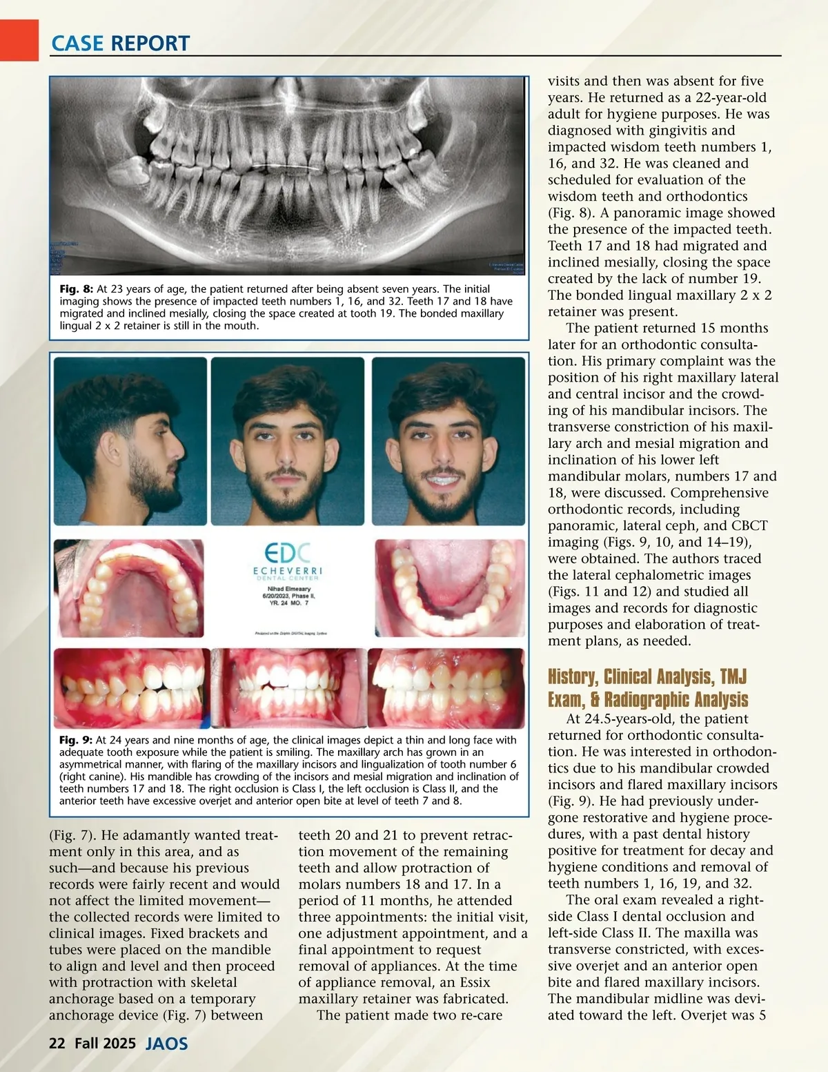
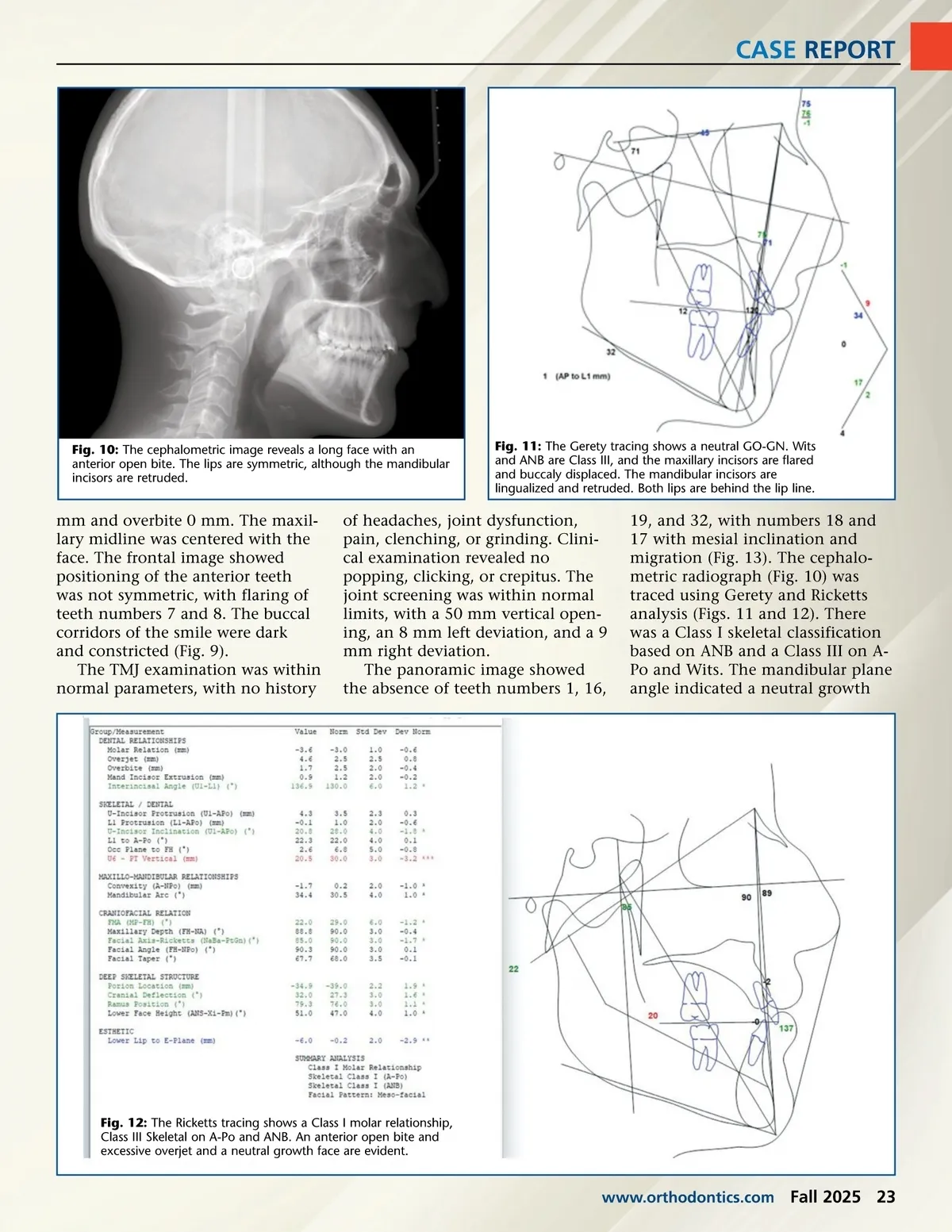
CASE REPORT Fig. 10: The cephalometric image reveals a long face with an anterior open bite. The lips are symmetric, although the mandibular incisors are retruded. Fig. 11: The Gerety tracing shows a neutral GO-GN. Wits and ANB are Class III, and the maxillary incisors are flared and buccaly displaced. The mandibular incisors are lingualized and retruded. Both lips are behind the lip line. mm and overbite 0 mm. The maxil-lary midline was centered with the face. The frontal image showed positioning of the anterior teeth was not symmetric, with flaring of teeth numbers 7 and 8. The buccal corridors of the smile were dark and constricted (Fig. 9). The TMJ examination was within normal parameters, with no history of headaches, joint dysfunction, pain, clenching, or grinding. Clini-cal examination revealed no popping, clicking, or crepitus. The joint screening was within normal limits, with a 50 mm vertical open-ing, an 8 mm left deviation, and a 9 mm right deviation. The panoramic image showed the absence of teeth numbers 1, 16, 19, and 32, with numbers 18 and 17 with mesial inclination and migration (Fig. 13). The cephalo-metric radiograph (Fig. 10) was traced using Gerety and Ricketts analysis (Figs. 11 and 12). There was a Class I skeletal classification based on ANB and a Class III on A-Po and Wits. The mandibular plane angle indicated a neutral growth Fig. 12: The Ricketts tracing shows a Class I molar relationship, Class III Skeletal on A-Po and ANB. An anterior open bite and excessive overjet and a neutral growth face are evident. www.orthodontics.com Fall 2025 23
Journal of the American Orthodontic Society Fall 2025: Page 23