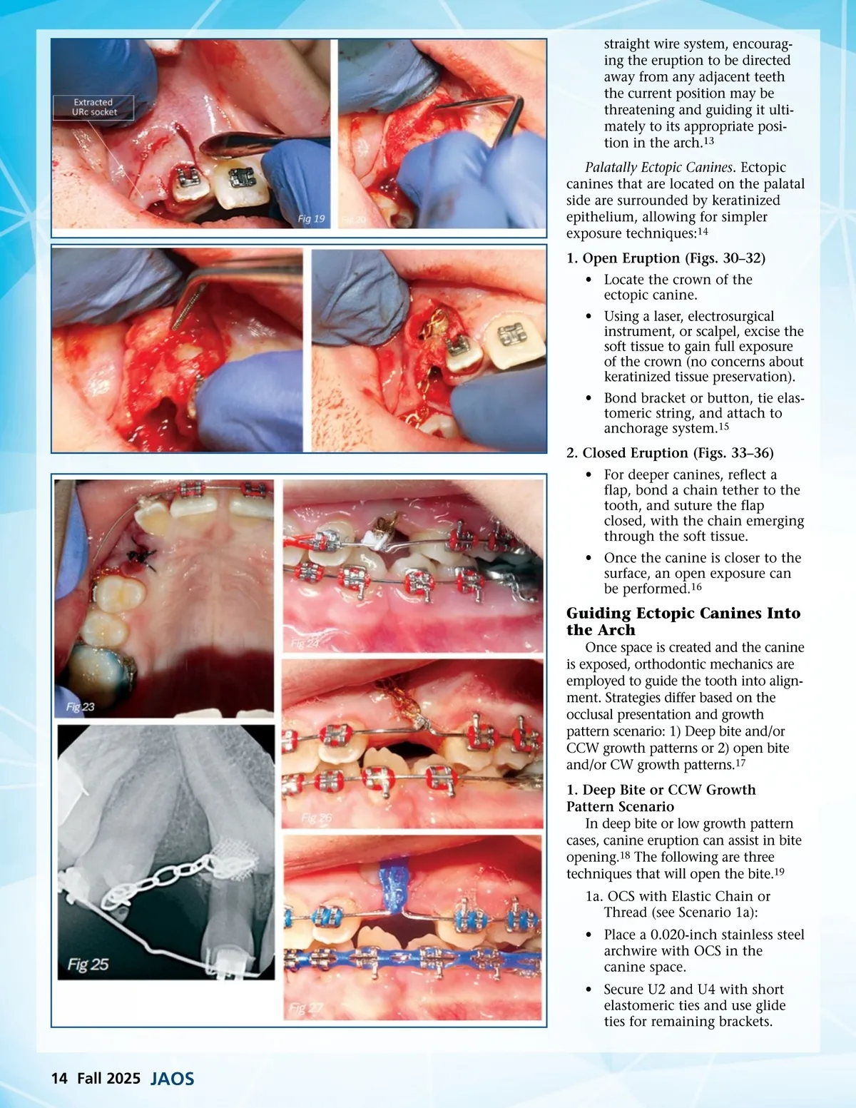
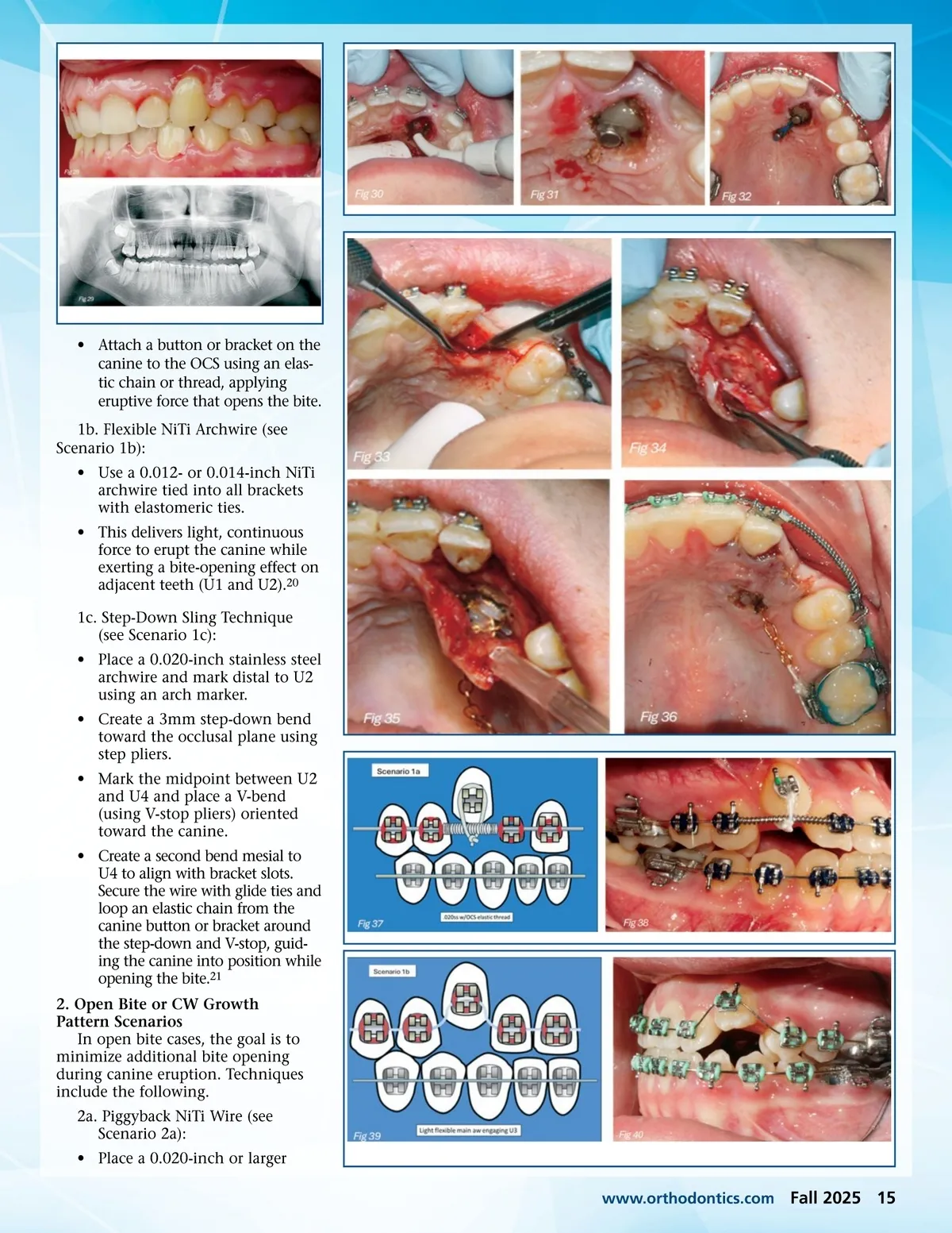
straight wire system, encourag-ing the eruption to be directed away from any adjacent teeth the current position may be threatening and guiding it ulti-mately to its appropriate posi-tion in the arch. 13 Palatally Ectopic Canines. Ectopic canines that are located on the palatal side are surrounded by keratinized epithelium, allowing for simpler exposure techniques: 14 1. Open Eruption (Figs. 30–32) • Locate the crown of the ectopic canine. • Using a laser, electrosurgical instrument, or scalpel, excise the soft tissue to gain full exposure of the crown (no concerns about keratinized tissue preservation). • Bond bracket or button, tie elas-tomeric string, and attach to anchorage system. 15 2. Closed Eruption (Figs. 33–36) • For deeper canines, reflect a flap, bond a chain tether to the tooth, and suture the flap closed, with the chain emerging through the soft tissue. • Once the canine is closer to the surface, an open exposure can be performed. 16 Guiding Ectopic Canines Into the Arch Once space is created and the canine is exposed, orthodontic mechanics are employed to guide the tooth into align-ment. Strategies differ based on the occlusal presentation and growth pattern scenario: 1) Deep bite and/or CCW growth patterns or 2) open bite and/or CW growth patterns. 17 1. Deep Bite or CCW Growth Pattern Scenario In deep bite or low growth pattern cases, canine eruption can assist in bite opening. 18 The following are three techniques that will open the bite. 19 1a. OCS with Elastic Chain or Thread (see Scenario 1a): • Place a 0.020-inch stainless steel archwire with OCS in the canine space. • Secure U2 and U4 with short elastomeric ties and use glide ties for remaining brackets. 14 Fall 2025 JAOS
Journal of the American Orthodontic Society Fall 2025: Page 14