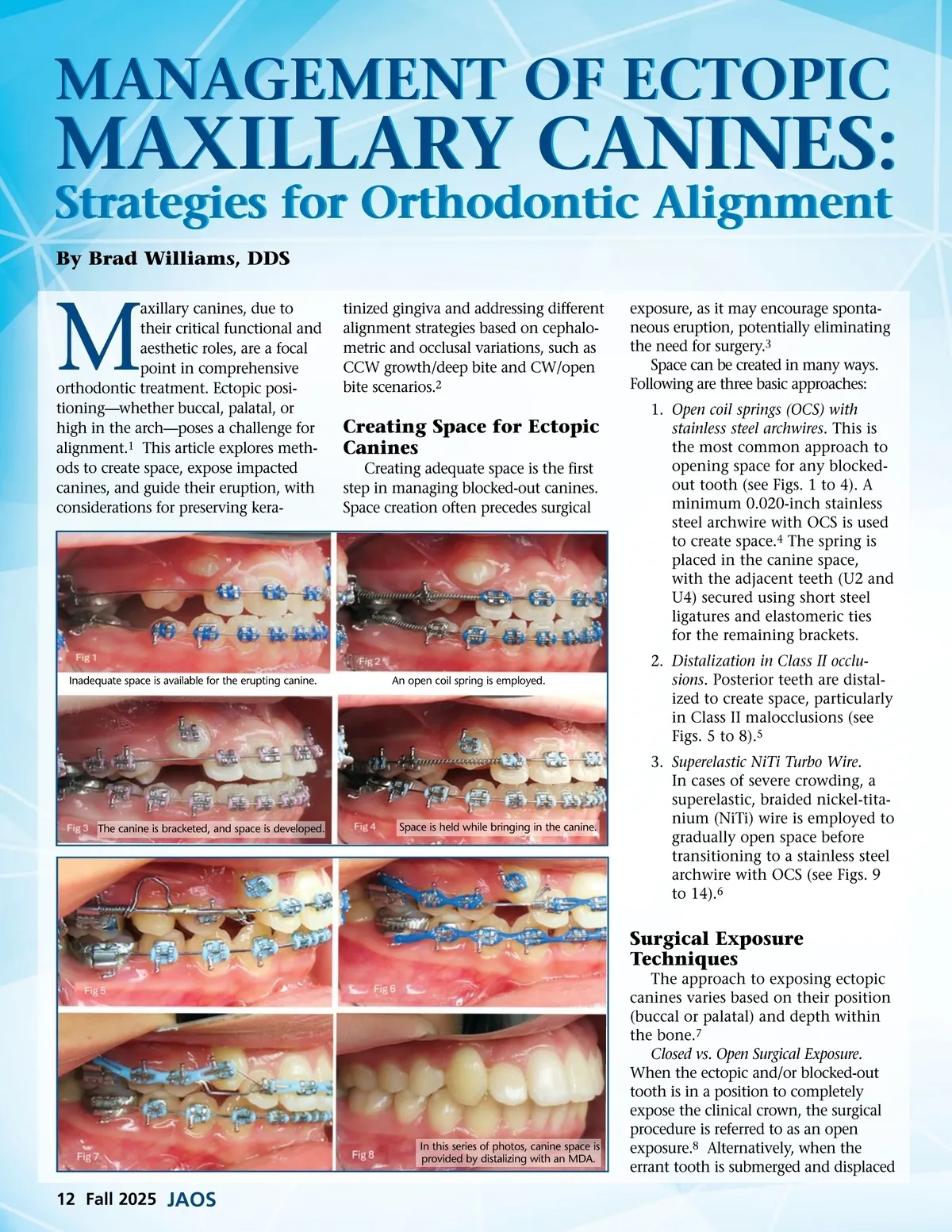
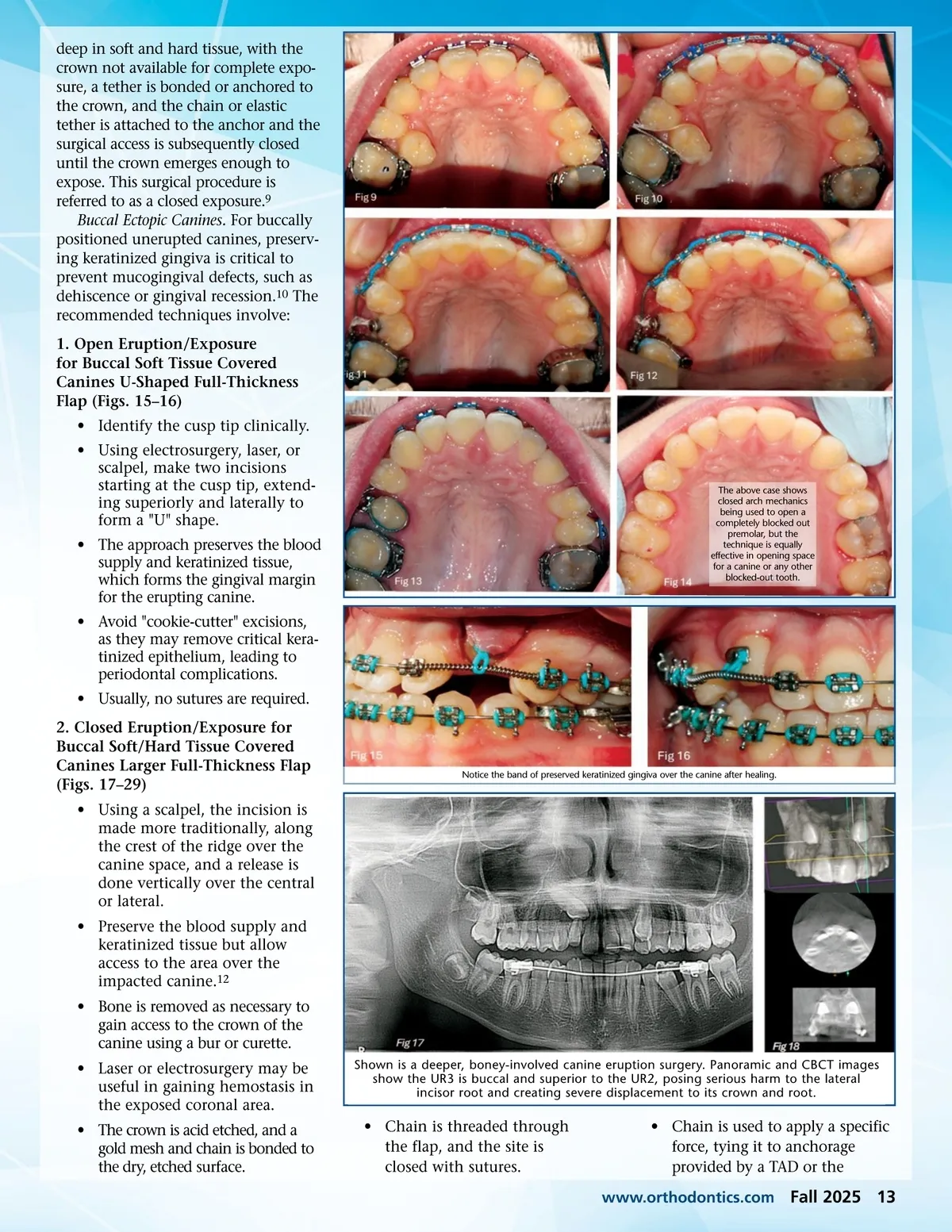
deep in soft and hard tissue, with the crown not available for complete expo-sure, a tether is bonded or anchored to the crown, and the chain or elastic tether is attached to the anchor and the surgical access is subsequently closed until the crown emerges enough to expose. This surgical procedure is referred to as a closed exposure. 9 Buccal Ectopic Canines. For buccally positioned unerupted canines, preserv-ing keratinized gingiva is critical to prevent mucogingival defects, such as dehiscence or gingival recession. 10 The recommended techniques involve: 1. Open Eruption/Exposure for Buccal Soft Tissue Covered Canines U-Shaped Full-Thickness Flap (Figs. 15–16) • Identify the cusp tip clinically. • Using electrosurgery, laser, or scalpel, make two incisions starting at the cusp tip, extend-ing superiorly and laterally to form a "U" shape. • The approach preserves the blood supply and keratinized tissue, which forms the gingival margin for the erupting canine. • Avoid "cookie-cutter" excisions, as they may remove critical kera-tinized epithelium, leading to periodontal complications. • Usually, no sutures are required. 2. Closed Eruption/Exposure for Buccal Soft/Hard Tissue Covered Canines Larger Full-Thickness Flap (Figs. 17–29) • Using a scalpel, the incision is made more traditionally, along the crest of the ridge over the canine space, and a release is done vertically over the central or lateral. • Preserve the blood supply and keratinized tissue but allow access to the area over the impacted canine. 12 • Bone is removed as necessary to gain access to the crown of the canine using a bur or curette. • Laser or electrosurgery may be useful in gaining hemostasis in the exposed coronal area. • The crown is acid etched, and a gold mesh and chain is bonded to the dry, etched surface. Shown is a deeper, boney-involved canine eruption surgery. Panoramic and CBCT images show the UR3 is buccal and superior to the UR2, posing serious harm to the lateral incisor root and creating severe displacement to its crown and root. The above case shows closed arch mechanics being used to open a completely blocked out premolar, but the technique is equally effective in opening space for a canine or any other blocked-out tooth. Notice the band of preserved keratinized gingiva over the canine after healing. • Chain is threaded through the flap, and the site is closed with sutures. • Chain is used to apply a specific force, tying it to anchorage provided by a TAD or the www.orthodontics.com Fall 2025 13
Journal of the American Orthodontic Society Fall 2025: Page 13