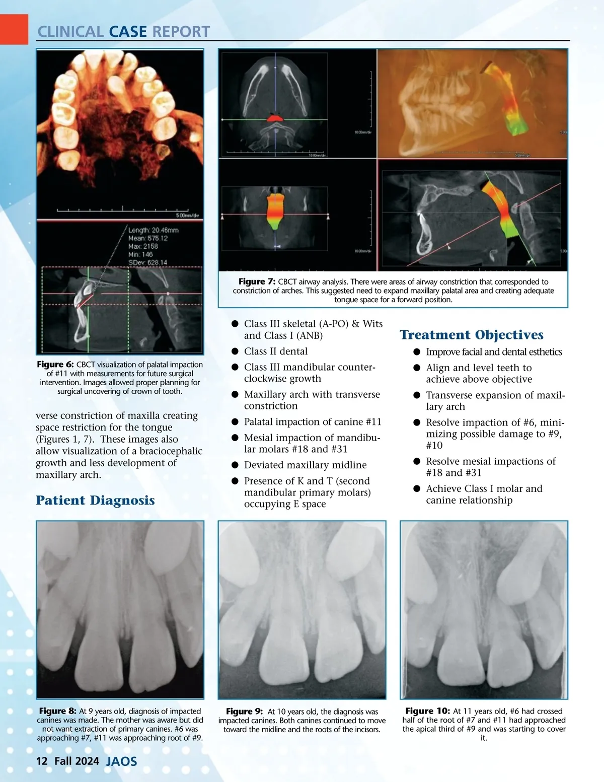
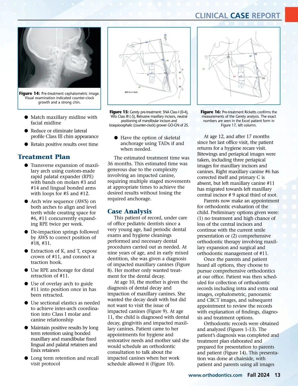
CLINICAL CASE REPORT Figure 7: CBCT airway analysis. There were areas of airway constriction that corresponded to constriction of arches. This suggested need to expand maxillary palatal area and creating adequate tongue space for a forward position. b Class III skeletal (A-PO) & Wits and Class I (ANB) b Class II dental Figure 6: CBCT visualization of palatal impaction of #11 with measurements for future surgical intervention. Images allowed proper planning for surgical uncovering of crown of tooth. Treatment Objectives b Improve facial and dental esthetics b Align and level teeth to achieve above objective b Transverse expansion of maxil-lary arch b Resolve impaction of #6, mini-mizing possible damage to #9, #10 b Resolve mesial impactions of #18 and #31 b Achieve Class I molar and canine relationship b Class III mandibular counter-clockwise growth b Maxillary arch with transverse constriction b Palatal impaction of canine #11 b Mesial impaction of mandibu-lar molars #18 and #31 b Deviated maxillary midline b Presence of K and T (second mandibular primary molars) occupying E space verse constriction of maxilla creating space restriction for the tongue (Figures 1, 7). These images also allow visualization of a braciocephalic growth and less development of maxillary arch. Patient Diagnosis Figure 8: At 9 years old, diagnosis of impacted canines was made. The mother was aware but did not want extraction of primary canines. #6 was approaching #7, #11 was approaching root of #9. Figure 9: At 10 years old, the diagnosis was impacted canines. Both canines continued to move toward the midline and the roots of the incisors. Figure 10: At 11 years old, #6 had crossed half of the root of #7 and #11 had approached the apical third of #9 and was starting to cover it. 12 Fall 2024 JAOS
Journal of the American Orthodontic Society Fall 2024: Page 12