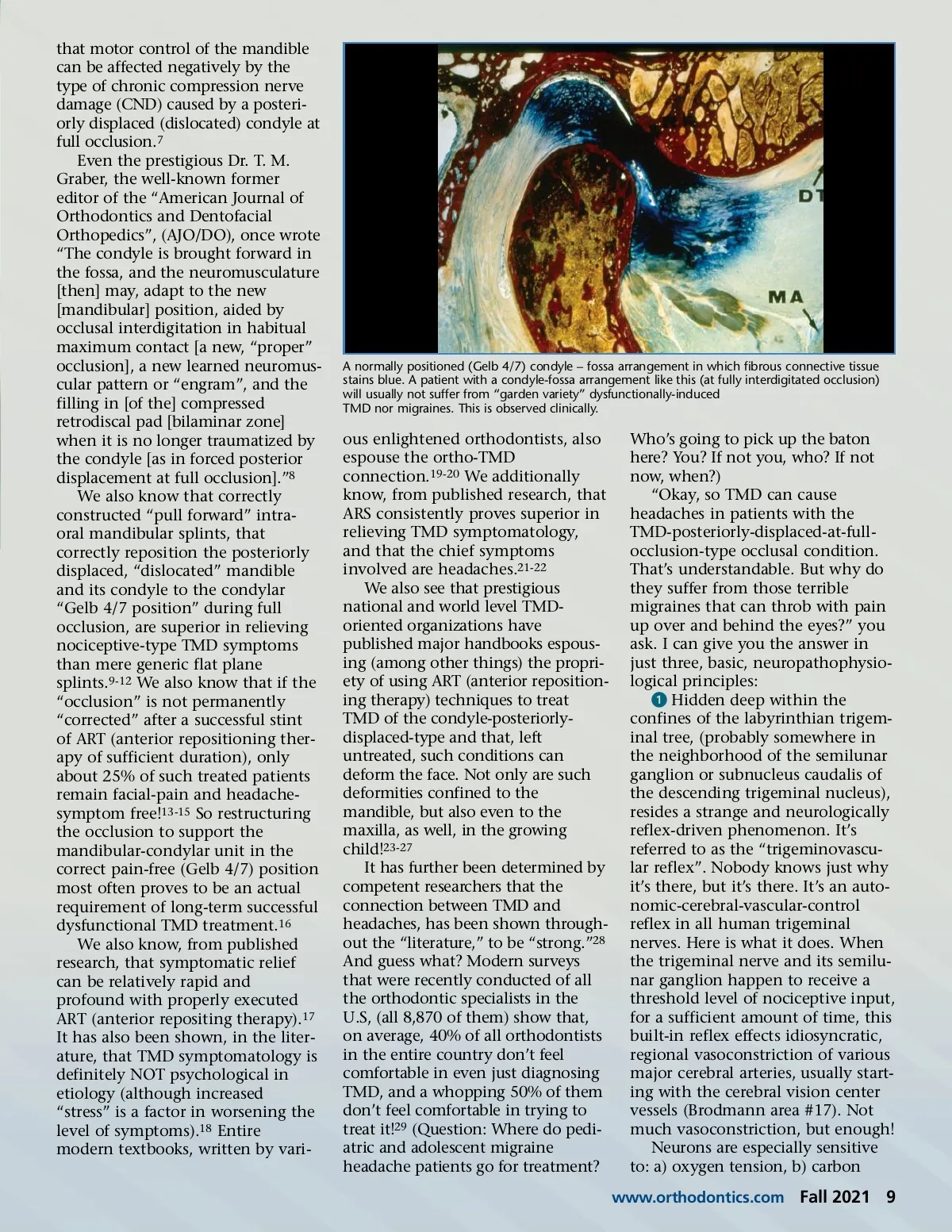
that motor control of the mandible can be affected negatively by the type of chronic compression nerve damage (CND) caused by a posteri-orly displaced (dislocated) condyle at full occlusion. 7 Even the prestigious Dr. T. M. Graber, the well-known former editor of the “American Journal of Orthodontics and Dentofacial Orthopedics”, (AJO/DO), once wrote “The condyle is brought forward in the fossa, and the neuromusculature [then] may, adapt to the new [mandibular] position, aided by occlusal interdigitation in habitual maximum contact [a new, “proper” occlusion], a new learned neuromus-cular pattern or “engram”, and the filling in [of the] compressed retrodiscal pad [bilaminar zone] when it is no longer traumatized by the condyle [as in forced posterior displacement at full occlusion].” 8 We also know that correctly constructed “pull forward” intra-oral mandibular splints, that correctly reposition the posteriorly displaced, “dislocated” mandible and its condyle to the condylar “Gelb 4/7 position” during full occlusion, are superior in relieving nociceptive-type TMD symptoms than mere generic flat plane splints. 9-12 We also know that if the “occlusion” is not permanently “corrected” after a successful stint of ART (anterior repositioning ther-apy of sufficient duration), only about 25% of such treated patients remain facial-pain and headache-symptom free! 13-15 So restructuring the occlusion to support the mandibular-condylar unit in the correct pain-free (Gelb 4/7) position most often proves to be an actual requirement of long-term successful dysfunctional TMD treatment. 16 We also know, from published research, that symptomatic relief can be relatively rapid and profound with properly executed ART (anterior repositing therapy). 17 It has also been shown, in the liter-ature, that TMD symptomatology is definitely NOT psychological in etiology (although increased “stress” is a factor in worsening the level of symptoms). 18 Entire modern textbooks, written by vari-A normally positioned (Gelb 4/7) condyle – fossa arrangement in which fibrous connective tissue stains blue. A patient with a condyle-fossa arrangement like this (at fully interdigitated occlusion) will usually not suffer from “garden variety” dysfunctionally-induced TMD nor migraines. This is observed clinically. ous enlightened orthodontists, also espouse the ortho-TMD connection. 19-20 We additionally know, from published research, that ARS consistently proves superior in relieving TMD symptomatology, and that the chief symptoms involved are headaches. 21-22 We also see that prestigious national and world level TMD-oriented organizations have published major handbooks espous-ing (among other things) the propri-ety of using ART (anterior reposition-ing therapy) techniques to treat TMD of the condyle-posteriorly-displaced-type and that, left untreated, such conditions can deform the face. Not only are such deformities confined to the mandible, but also even to the maxilla, as well, in the growing child! 23-27 It has further been determined by competent researchers that the connection between TMD and headaches, has been shown through-out the “literature,” to be “strong.” 28 And guess what? Modern surveys that were recently conducted of all the orthodontic specialists in the U.S, (all 8,870 of them) show that, on average, 40% of all orthodontists in the entire country don’t feel comfortable in even just diagnosing TMD, and a whopping 50% of them don’t feel comfortable in trying to treat it! 29 (Question: Where do pedi-atric and adolescent migraine headache patients go for treatment? Who’s going to pick up the baton here? You? If not you, who? If not now, when?) “Okay, so TMD can cause headaches in patients with the TMD-posteriorly-displaced-at-full-occlusion-type occlusal condition. That’s understandable. But why do they suffer from those terrible migraines that can throb with pain up over and behind the eyes?” you ask. I can give you the answer in just three, basic, neuropathophysio-logical principles: ᕡ Hidden deep within the confines of the labyrinthian trigem-inal tree, (probably somewhere in the neighborhood of the semilunar ganglion or subnucleus caudalis of the descending trigeminal nucleus), resides a strange and neurologically reflex-driven phenomenon. It’s referred to as the “trigeminovascu-lar reflex”. Nobody knows just why it’s there, but it’s there. It’s an auto-nomic-cerebral-vascular-control reflex in all human trigeminal nerves. Here is what it does. When the trigeminal nerve and its semilu-nar ganglion happen to receive a threshold level of nociceptive input, for a sufficient amount of time, this built-in reflex effects idiosyncratic, regional vasoconstriction of various major cerebral arteries, usually start-ing with the cerebral vision center vessels (Brodmann area #17). Not much vasoconstriction, but enough! Neurons are especially sensitive to: a) oxygen tension, b) carbon www.orthodontics.com Fall 2021 9
Journal of the American Orthodontic Society Fall 2021: Page 9