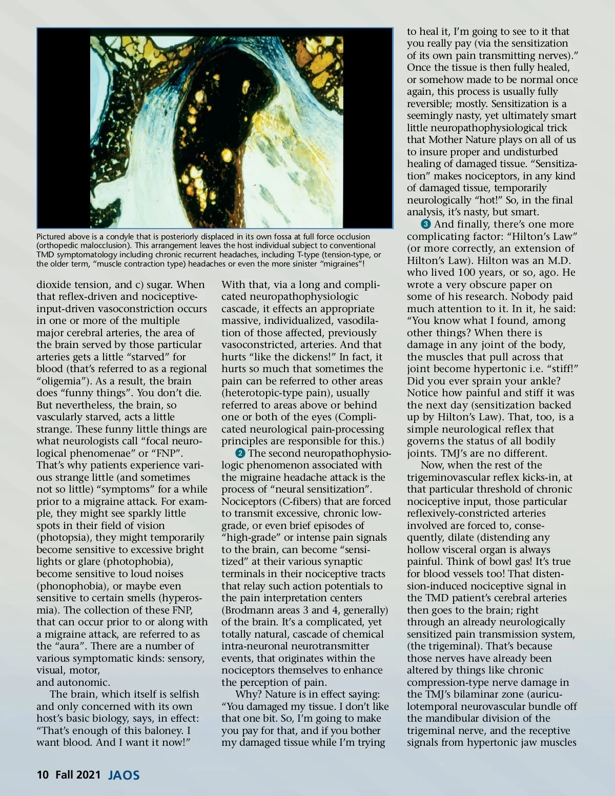
Pictured above is a condyle that is posteriorly displaced in its own fossa at full force occlusion (orthopedic malocclusion). This arrangement leaves the host individual subject to conventional TMD symptomatology including chronic recurrent headaches, including T-type (tension-type, or the older term, “muscle contraction type) headaches or even the more sinister “migraines”! dioxide tension, and c) sugar. When that reflex-driven and nociceptive-input-driven vasoconstriction occurs in one or more of the multiple major cerebral arteries, the area of the brain served by those particular arteries gets a little “starved” for blood (that’s referred to as a regional “oligemia”). As a result, the brain does “funny things”. You don’t die. But nevertheless, the brain, so vascularly starved, acts a little strange. These funny little things are what neurologists call “focal neuro-logical phenomenae” or “FNP”. That’s why patients experience vari-ous strange little (and sometimes not so little) “symptoms” for a while prior to a migraine attack. For exam-ple, they might see sparkly little spots in their field of vision (photopsia), they might temporarily become sensitive to excessive bright lights or glare (photophobia), become sensitive to loud noises (phonophobia), or maybe even sensitive to certain smells (hyperos-mia). The collection of these FNP, that can occur prior to or along with a migraine attack, are referred to as the “aura”. There are a number of various symptomatic kinds: sensory, visual, motor, and autonomic. The brain, which itself is selfish and only concerned with its own host’s basic biology, says, in effect: “That’s enough of this baloney. I want blood. And I want it now!” With that, via a long and compli-cated neuropathophysiologic cascade, it effects an appropriate massive, individualized, vasodila-tion of those affected, previously vasoconstricted, arteries. And that hurts “like the dickens!” In fact, it hurts so much that sometimes the pain can be referred to other areas (heterotopic-type pain), usually referred to areas above or behind one or both of the eyes (Compli-cated neurological pain-processing principles are responsible for this.) ᕢ The second neuropathophysio-logic phenomenon associated with the migraine headache attack is the process of “neural sensitization”. Nociceptors (C-fibers) that are forced to transmit excessive, chronic low-grade, or even brief episodes of “high-grade” or intense pain signals to the brain, can become “sensi-tized” at their various synaptic terminals in their nociceptive tracts that relay such action potentials to the pain interpretation centers (Brodmann areas 3 and 4, generally) of the brain. It’s a complicated, yet totally natural, cascade of chemical intra-neuronal neurotransmitter events, that originates within the nociceptors themselves to enhance the perception of pain. Why? Nature is in effect saying: “You damaged my tissue. I don’t like that one bit. So, I’m going to make you pay for that, and if you bother my damaged tissue while I’m trying to heal it, I’m going to see to it that you really pay (via the sensitization of its own pain transmitting nerves).” Once the tissue is then fully healed, or somehow made to be normal once again, this process is usually fully reversible; mostly. Sensitization is a seemingly nasty, yet ultimately smart little neuropathophysiological trick that Mother Nature plays on all of us to insure proper and undisturbed healing of damaged tissue. “Sensitiza-tion” makes nociceptors, in any kind of damaged tissue, temporarily neurologically “hot!” So, in the final analysis, it’s nasty, but smart. ᕣ And finally, there’s one more complicating factor: “Hilton’s Law” (or more correctly, an extension of Hilton’s Law). Hilton was an M.D. who lived 100 years, or so, ago. He wrote a very obscure paper on some of his research. Nobody paid much attention to it. In it, he said: “You know what I found, among other things? When there is damage in any joint of the body, the muscles that pull across that joint become hypertonic i.e. “stiff!” Did you ever sprain your ankle? Notice how painful and stiff it was the next day (sensitization backed up by Hilton’s Law). That, too, is a simple neurological reflex that governs the status of all bodily joints. TMJ’s are no different. Now, when the rest of the trigeminovascular reflex kicks-in, at that particular threshold of chronic nociceptive input, those particular reflexively-constricted arteries involved are forced to, conse-quently, dilate (distending any hollow visceral organ is always painful. Think of bowl gas! It’s true for blood vessels too! That disten-sion-induced nociceptive signal in the TMD patient’s cerebral arteries then goes to the brain; right through an already neurologically sensitized pain transmission system, (the trigeminal). That’s because those nerves have already been altered by things like chronic compression-type nerve damage in the TMJ’s bilaminar zone (auricu-lotemporal neurovascular bundle off the mandibular division of the trigeminal nerve, and the receptive signals from hypertonic jaw muscles 10 Fall 2021 JAOS
Journal of the American Orthodontic Society Fall 2021: Page 10