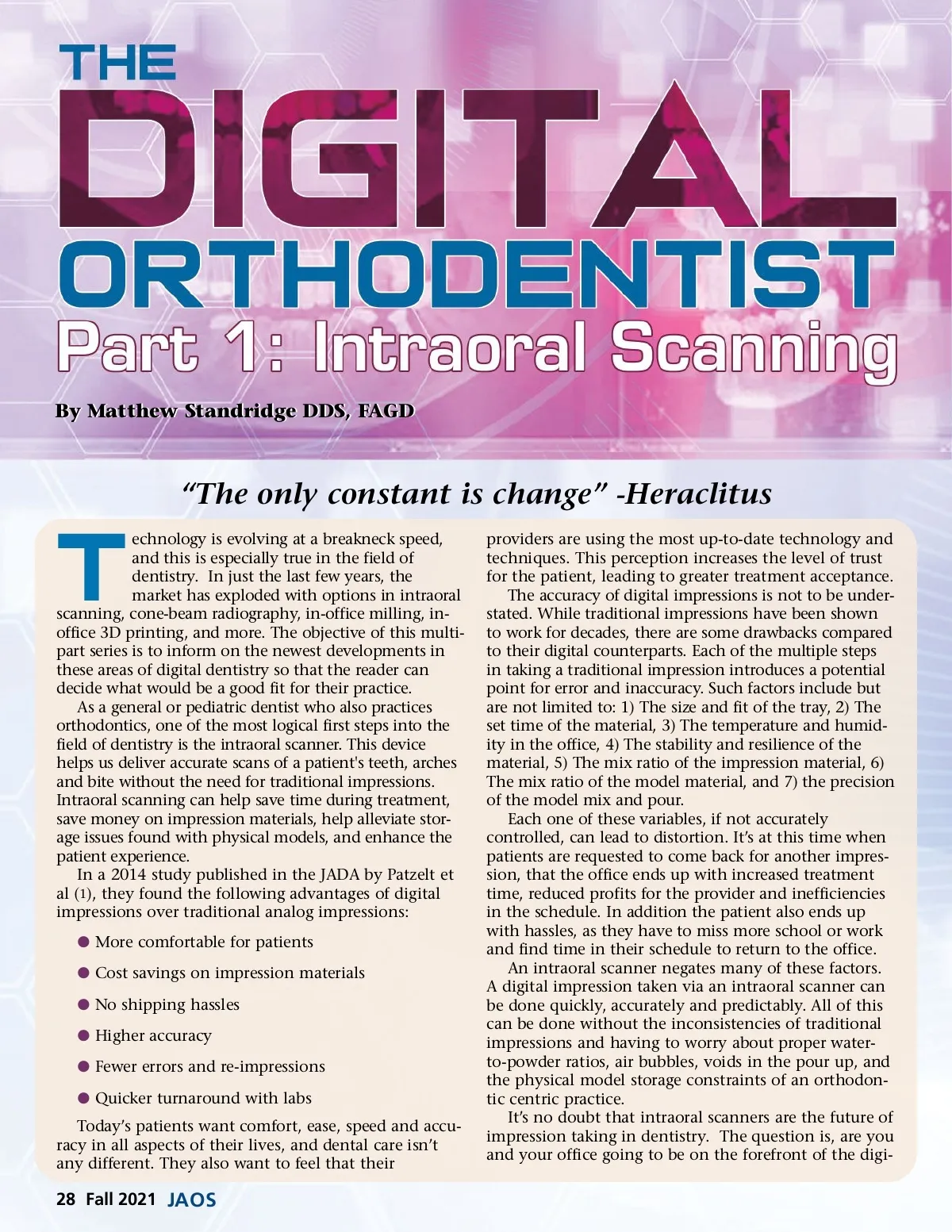
By Matthew Standridge DDS, FAGD “The only constant is change” -Heraclitus T echnology is evolving at a breakneck speed, and this is especially true in the field of dentistry. In just the last few years, the market has exploded with options in intraoral scanning, cone-beam radiography, in-office milling, in-office 3D printing, and more. The objective of this multi-part series is to inform on the newest developments in these areas of digital dentistry so that the reader can decide what would be a good fit for their practice. As a general or pediatric dentist who also practices orthodontics, one of the most logical first steps into the field of dentistry is the intraoral scanner. This device helps us deliver accurate scans of a patient's teeth, arches and bite without the need for traditional impressions. Intraoral scanning can help save time during treatment, save money on impression materials, help alleviate stor-age issues found with physical models, and enhance the patient experience. In a 2014 study published in the JADA by Patzelt et al ( 1 ), they found the following advantages of digital impressions over traditional analog impressions: b More comfortable for patients b Cost savings on impression materials b No shipping hassles b Higher accuracy b Fewer errors and re-impressions b Quicker turnaround with labs Today’s patients want comfort, ease, speed and accu-racy in all aspects of their lives, and dental care isn’t any different. They also want to feel that their providers are using the most up-to-date technology and techniques. This perception increases the level of trust for the patient, leading to greater treatment acceptance. The accuracy of digital impressions is not to be under-stated. While traditional impressions have been shown to work for decades, there are some drawbacks compared to their digital counterparts. Each of the multiple steps in taking a traditional impression introduces a potential point for error and inaccuracy. Such factors include but are not limited to: 1) The size and fit of the tray, 2) The set time of the material, 3) The temperature and humid-ity in the office, 4) The stability and resilience of the material, 5) The mix ratio of the impression material, 6) The mix ratio of the model material, and 7) the precision of the model mix and pour. Each one of these variables, if not accurately controlled, can lead to distortion. It’s at this time when patients are requested to come back for another impres-sion, that the office ends up with increased treatment time, reduced profits for the provider and inefficiencies in the schedule. In addition the patient also ends up with hassles, as they have to miss more school or work and find time in their schedule to return to the office. An intraoral scanner negates many of these factors. A digital impression taken via an intraoral scanner can be done quickly, accurately and predictably. All of this can be done without the inconsistencies of traditional impressions and having to worry about proper water-to-powder ratios, air bubbles, voids in the pour up, and the physical model storage constraints of an orthodon-tic centric practice. It’s no doubt that intraoral scanners are the future of impression taking in dentistry. The question is, are you and your office going to be on the forefront of the digi-28 Fall 2021 JAOS
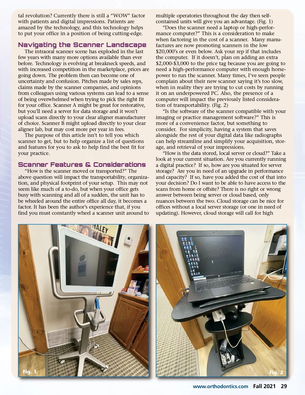
Journal of the American Orthodontic Society Fall 2021: Page 28