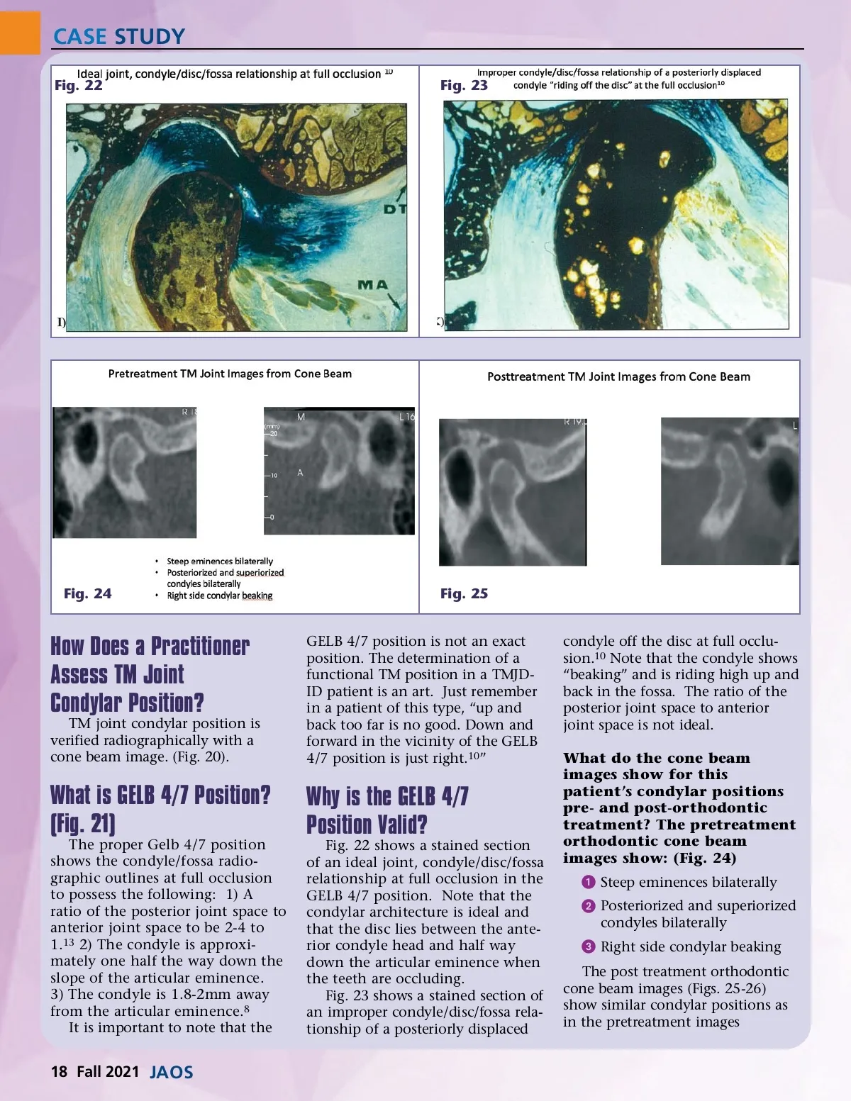
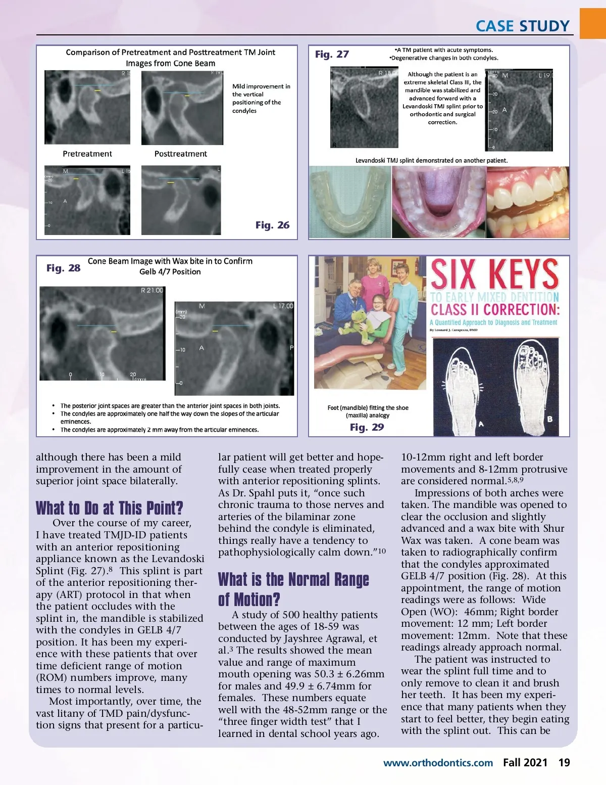
CASE STUDY Fig. 22 Fig. 23 Fig. 24 Fig. 25 GELB 4/7 position is not an exact position. The determination of a functional TM position in a TMJD-ID patient is an art. Just remember in a patient of this type, “up and back too far is no good. Down and forward in the vicinity of the GELB 4/7 position is just right. 10 ” condyle off the disc at full occlu-sion. 10 Note that the condyle shows “beaking” and is riding high up and back in the fossa. The ratio of the posterior joint space to anterior joint space is not ideal. What do the cone beam images show for this patient’s condylar positions pre-and post-orthodontic treatment? The pretreatment orthodontic cone beam images show: (Fig. 24) ᕡ Steep eminences bilaterally ᕢ Posteriorized and superiorized condyles bilaterally ᕣ Right side condylar beaking The post treatment orthodontic cone beam images (Figs. 25-26) show similar condylar positions as in the pretreatment images How Does a Practitioner Assess TM Joint Condylar Position? TM joint condylar position is verified radiographically with a cone beam image. (Fig. 20). What is GELB 4/7 Position? (Fig. 21) The proper Gelb 4/7 position shows the condyle/fossa radio-graphic outlines at full occlusion to possess the following: 1) A ratio of the posterior joint space to anterior joint space to be 2-4 to 1. 13 2) The condyle is approxi-mately one half the way down the slope of the articular eminence. 3) The condyle is 1.8-2mm away from the articular eminence. 8 It is important to note that the Why is the GELB 4/7 Position Valid? Fig. 22 shows a stained section of an ideal joint, condyle/disc/fossa relationship at full occlusion in the GELB 4/7 position. Note that the condylar architecture is ideal and that the disc lies between the ante-rior condyle head and half way down the articular eminence when the teeth are occluding. Fig. 23 shows a stained section of an improper condyle/disc/fossa rela-tionship of a posteriorly displaced 18 Fall 2021 JAOS
Journal of the American Orthodontic Society Fall 2021: Page 18