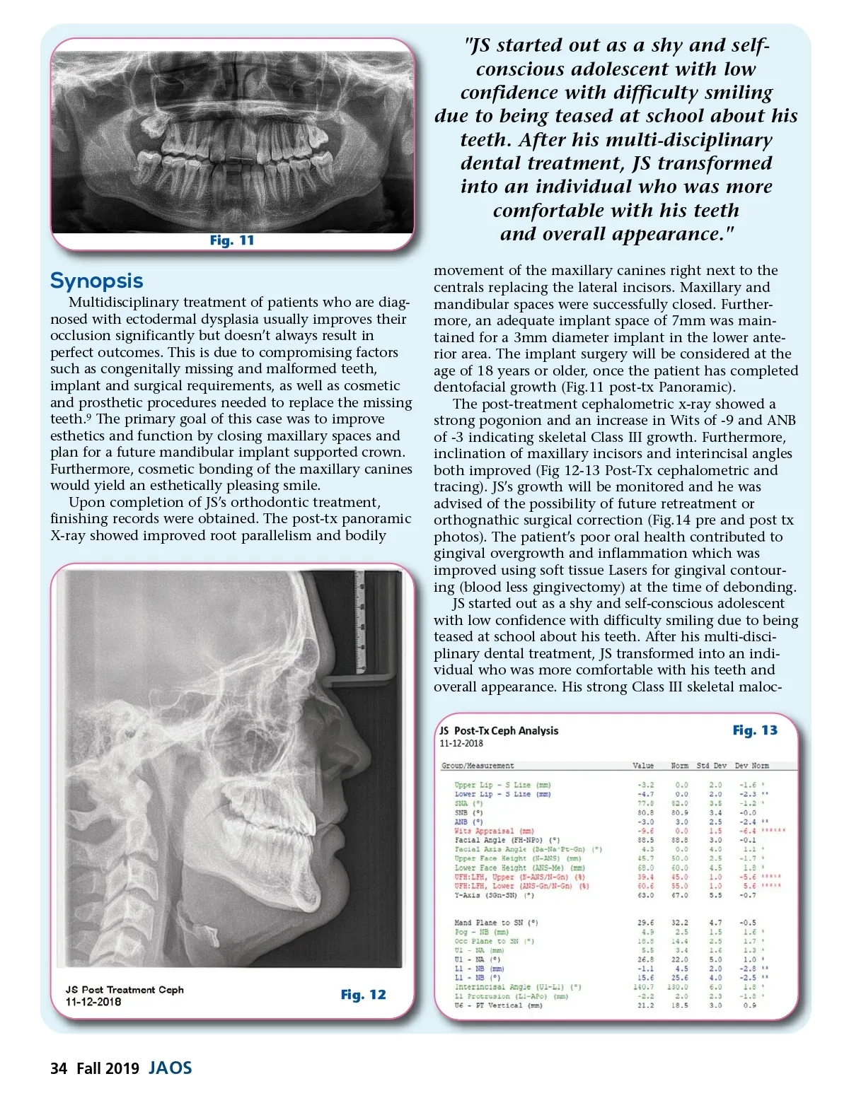
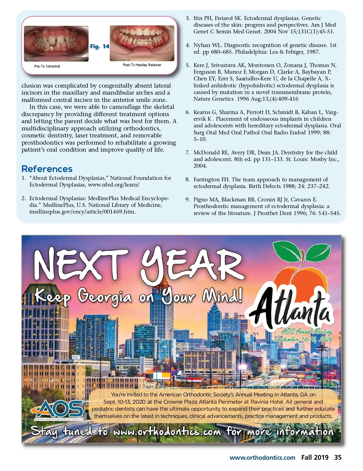
Fig. 11 "JS started out as a shy and self-conscious adolescent with low confidence with difficulty smiling due to being teased at school about his teeth. After his multi-disciplinary dental treatment, JS transformed into an individual who was more comfortable with his teeth and overall appearance." movement of the maxillary canines right next to the centrals replacing the lateral incisors. Maxillary and mandibular spaces were successfully closed. Further-more, an adequate implant space of 7mm was main-tained for a 3mm diameter implant in the lower ante-rior area. The implant surgery will be considered at the age of 18 years or older, once the patient has completed dentofacial growth (Fig.11 post-tx Panoramic). The post-treatment cephalometric x-ray showed a strong pogonion and an increase in Wits of -9 and ANB of -3 indicating skeletal Class III growth. Furthermore, inclination of maxillary incisors and interincisal angles both improved (Fig 12-13 Post-Tx cephalometric and tracing). JS’s growth will be monitored and he was advised of the possibility of future retreatment or orthognathic surgical correction (Fig.14 pre and post tx photos). The patient’s poor oral health contributed to gingival overgrowth and inflammation which was improved using soft tissue Lasers for gingival contour-ing (blood less gingivectomy) at the time of debonding. JS started out as a shy and self-conscious adolescent with low confidence with difficulty smiling due to being teased at school about his teeth. After his multi-disci-plinary dental treatment, JS transformed into an indi-vidual who was more comfortable with his teeth and overall appearance. His strong Class III skeletal maloc-Fig. 13 Synopsis Multidisciplinary treatment of patients who are diag-nosed with ectodermal dysplasia usually improves their occlusion significantly but doesn’t always result in perfect outcomes. This is due to compromising factors such as congenitally missing and malformed teeth, implant and surgical requirements, as well as cosmetic and prosthetic procedures needed to replace the missing teeth. 9 The primary goal of this case was to improve esthetics and function by closing maxillary spaces and plan for a future mandibular implant supported crown. Furthermore, cosmetic bonding of the maxillary canines would yield an esthetically pleasing smile. Upon completion of JS’s orthodontic treatment, finishing records were obtained. The post-tx panoramic X-ray showed improved root parallelism and bodily Fig. 12 34 Fall 2019 JAOS
Journal of the American Orthodontic Society Fall 2019: Page 34