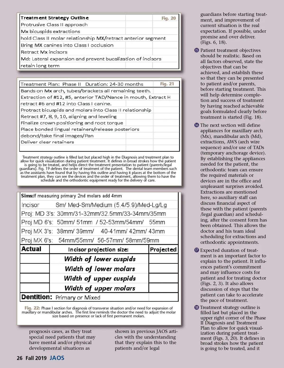
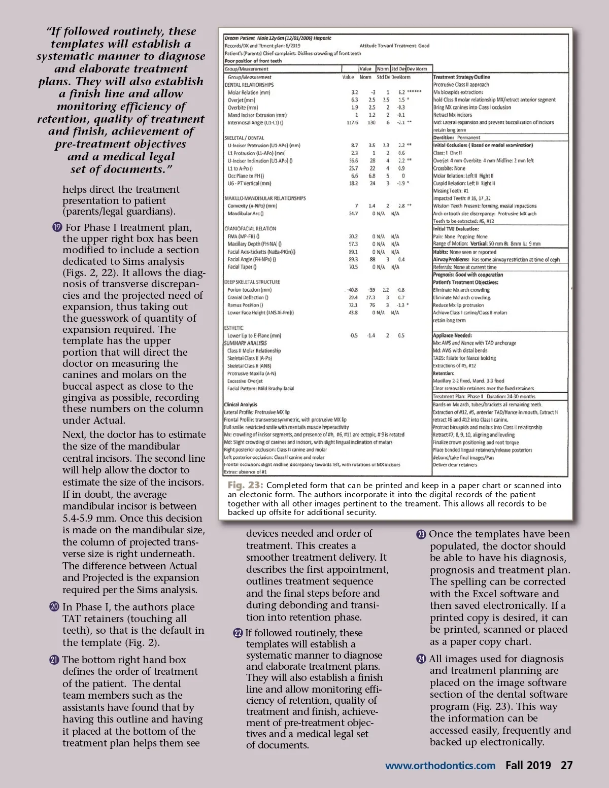
“If followed routinely, these templates will establish a systematic manner to diagnose and elaborate treatment plans. They will also establish a finish line and allow monitoring efficiency of retention, quality of treatment and finish, achievement of pre-treatment objectives and a medical legal set of documents.” helps direct the treatment presentation to patient (parents/legal guardians). ƾ For Phase I treatment plan, the upper right box has been modified to include a section dedicated to Sims analysis (Figs. 2, 22). It allows the diag-nosis of transverse discrepan-cies and the projected need of expansion, thus taking out the guesswork of quantity of expansion required. The template has the upper portion that will direct the doctor on measuring the canines and molars on the buccal aspect as close to the gingiva as possible, recording these numbers on the column under Actual. Next, the doctor has to estimate the size of the mandibular central incisors. The second line will help allow the doctor to estimate the size of the incisors. If in doubt, the average mandibular incisor is between 5.4-5.9 mm. Once this decision is made on the mandibular size, the column of projected trans-verse size is right underneath. The difference between Actual and Projected is the expansion required per the Sims analysis. ƿ In Phase I, the authors place TAT retainers (touching all teeth), so that is the default in the template (Fig. 2). © 21 The bottom right hand box defines the order of treatment of the patient. The dental team members such as the assistants have found that by having this outline and having it placed at the bottom of the treatment plan helps them see Fig. 23: Completed form that can be printed and keep in a paper chart or scanned into an electonic form. The authors incorporate it into the digital records of the patient together with all other images pertinent to the treament. This allows all records to be backed up offsite for additional security. devices needed and order of treatment. This creates a smoother treatment delivery. It describes the first appointment, outlines treatment sequence and the final steps before and during debonding and transi-tion into retention phase. © 22 If followed routinely, these templates will establish a systematic manner to diagnose and elaborate treatment plans. They will also establish a finish line and allow monitoring effi-ciency of retention, quality of treatment and finish, achieve-ment of pre-treatment objec-tives and a medical legal set of documents. © 23 Once the templates have been populated, the doctor should be able to have his diagnosis, prognosis and treatment plan. The spelling can be corrected with the Excel software and then saved electronically. If a printed copy is desired, it can be printed, scanned or placed as a paper copy chart. © 24 All images used for diagnosis and treatment planning are placed on the image software section of the dental software program (Fig. 23). This way the information can be accessed easily, frequently and backed up electronically. www.orthodontics.com Fall 2019 27
Journal of the American Orthodontic Society Fall 2019: Page 27