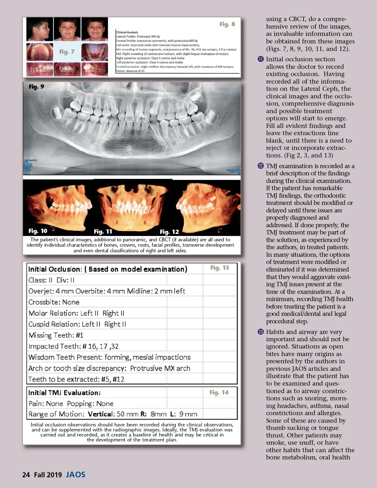
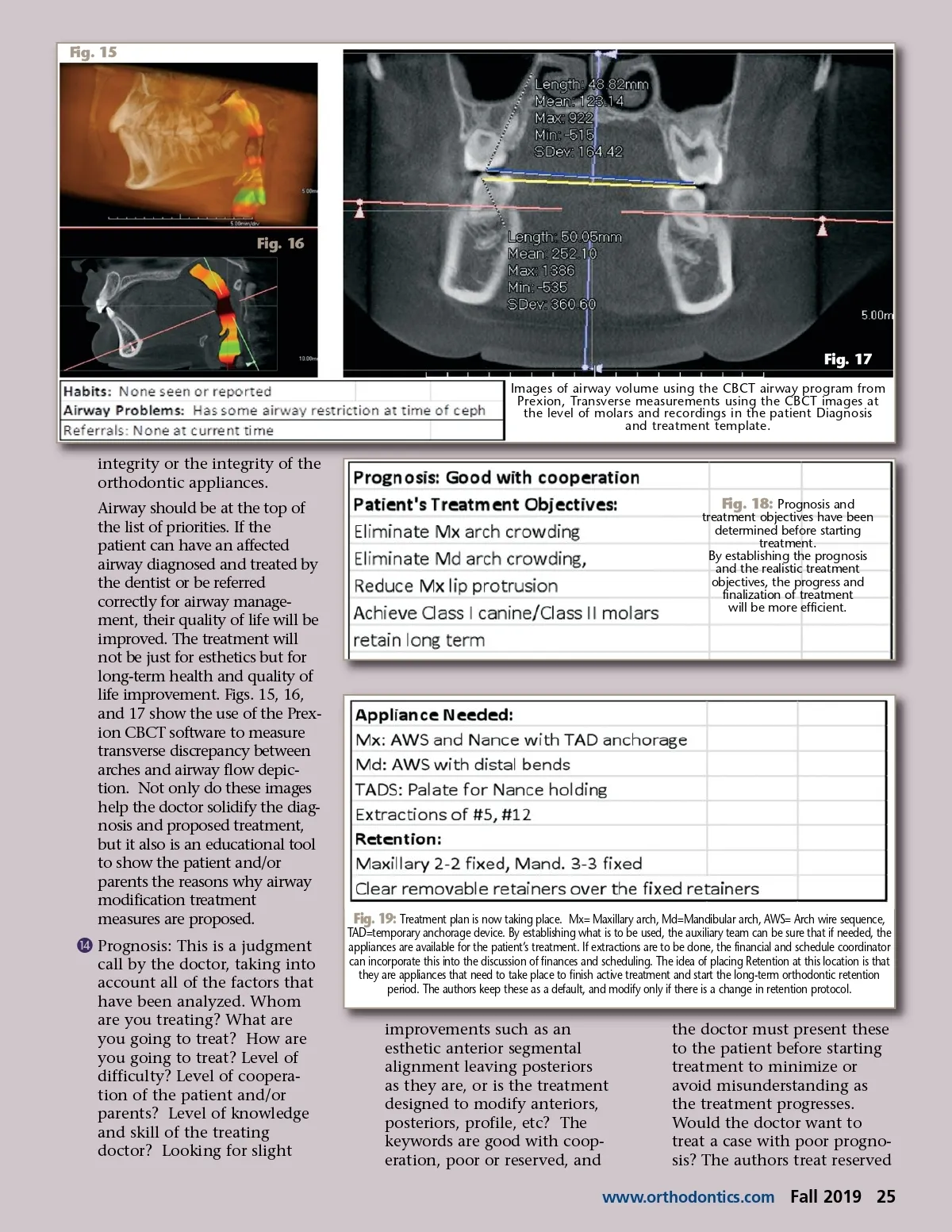
Fig. 8 Fig. 7 using a CBCT, do a compre-hensive review of the images, as invaluable information can be obtained from these images (Figs. 7, 8, 9, 10, 11, and 12). ¸ Initial occlusion section allows the doctor to record existing occlusion. Having recorded all of the informa-tion on the Lateral Ceph, the clinical images and the occlu-sion, comprehensive diagnosis and possible treatment options will start to emerge. Fill all evident findings and leave the extractions line blank, until there is a need to reject or incorporate extrac-tions. (Fig 2, 3, and 13) ¹ TMJ examination is recorded as a brief description of the findings during the clinical examination. If the patient has remarkable TMJ findings, the orthodontic treatment should be modified or delayed until these issues are properly diagnosed and addressed. If done properly, the TMJ treatment may be part of the solution, as experienced by the authors, in treated patients. In many situations, the options of treatment were modified or eliminated if it was determined that they would aggravate exist-ing TMJ issues present at the time of the examination. At a minimum, recording TMJ health before treating the patient is a good medical/dental and legal procedural step. Ƹ Habits and airway are very important and should not be ignored. Situations as open bites have many origins as presented by the authors in previous JAOS articles and illustrate that the patient has to be examined and ques-tioned as to airway constric-tions such as snoring, morn-ing headaches, asthma, nasal constrictions and allergies. Some of these are caused by thumb sucking or tongue thrust. Other patients may smoke, use snuff, or have other habits that can affect the bone metabolism, oral health Fig. 9 Fig. 10 Fig. 11 Fig. 12 The patient’s clinical images, additional to panoramic, and CBCT (if available) are all used to identify individual characteristics of bones, crowns, roots, facial profiles, transverse development and even dental classifications of right and left sides. Fig. 13 Fig. 14 Initial occlusion observations should have been recorded during the clinical observations, and can be supplemented with the radiographic images. Ideally, the TMJ evaluation was carried out and recorded, as it creates a baseline of health and may be critical in the development of the treatment plan. 24 Fall 2019 JAOS
Journal of the American Orthodontic Society Fall 2019: Page 24