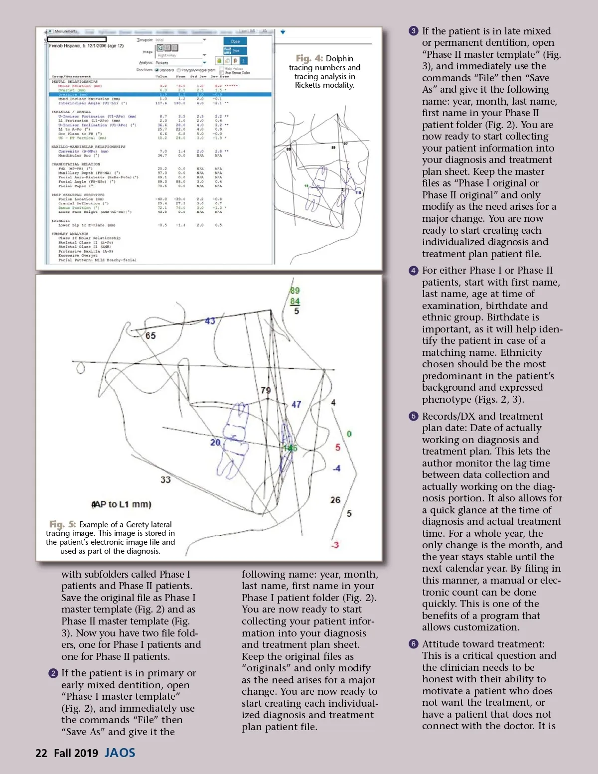
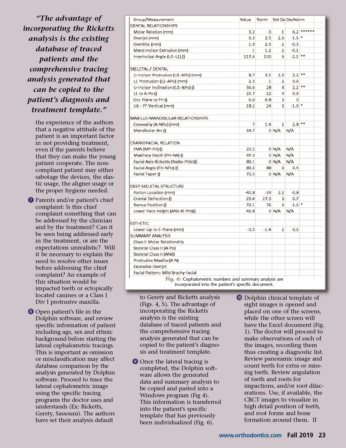
“The advantage of incorporating the Ricketts analysis is the existing database of traced patients and the comprehensive tracing analysis generated that can be copied to the patient’s diagnosis and treatment template.” the experience of the authors that a negative attitude of the patient is an important factor in not providing treatment, even if the parents believe that they can make the young patient cooperate. The non-compliant patient may either sabotage the devices, the elas-tic usage, the aligner usage or the proper hygiene needed. ᕧ Parents and/or patient’s chief complaint: Is this chief complaint something that can be addressed by the clinician and by the treatment? Can it be seen being addressed early in the treatment, or are the expectations unrealistic? Will it be necessary to explain the need to resolve other issues before addressing the chief complaint? An example of this situation would be impacted teeth or ectopically located canines or a Class I Div I protrusive maxilla. ᕨ Open patient’s file in the Dolphin software, and review specific information of patient including age, sex and ethnic background before starting the lateral cephalometric tracings. This is important as omission or misclassification may affect database comparison by the analysis generated by Dolphin software. Proceed to trace the lateral cephalometric image using the specific tracing programs the doctor uses and understands (Ex: Ricketts, Gerety, Sassouni). The authors have set their analysis default Fig. 6: Cephalometric numbers and summary analysis are incorporated into the patient’s specific document. to Gerety and Ricketts analysis (Figs. 4, 5). The advantage of incorporating the Ricketts analysis is the existing database of traced patients and the comprehensive tracing analysis generated that can be copied to the patient’s diagno-sis and treatment template. ᕩ Once the lateral tracing is completed, the Dolphin soft-ware allows the generated data and summary analysis to be copied and pasted into a Windows program (Fig 4). This information is transferred into the patient’s specific template that has previously been individualized (Fig. 6). µ Dolphin clinical template of eight images is opened and placed on one of the screens, while the other screen will have the Excel document (Fig. 1). The doctor will proceed to make observations of each of the images, recording them thus creating a diagnostic list. Review panoramic image and count teeth for extra or miss-ing teeth. Review angulation of teeth and roots for impactions, and/or root dilac-erations. Use, if available, the CBCT images to visualize in high detail position of teeth, and root forms and bone formation around them. If www.orthodontics.com Fall 2019 23
Journal of the American Orthodontic Society Fall 2019: Page 23