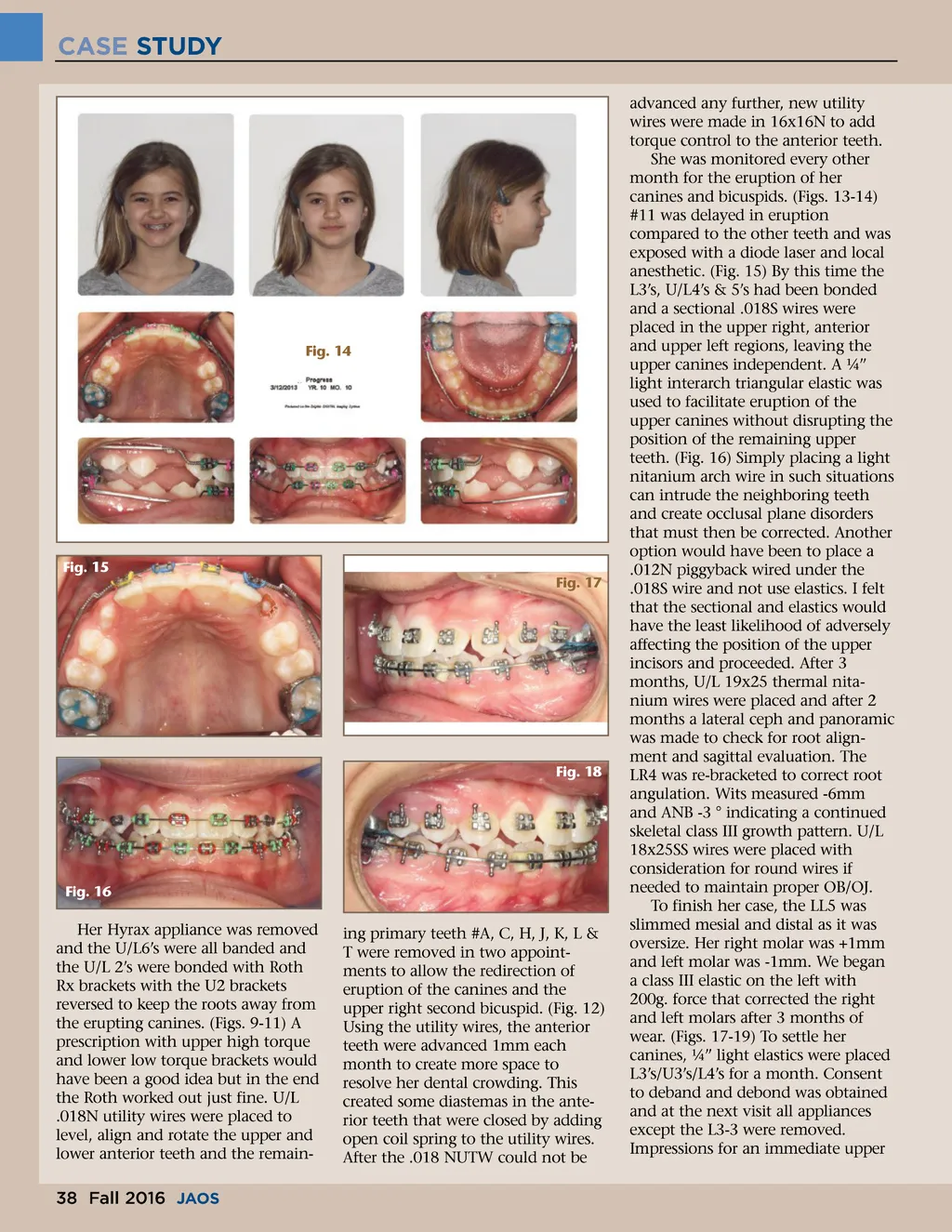
CASE STUDY advanced any further, new utility wires were made in 16x16N to add torque control to the anterior teeth. She was monitored every other month for the eruption of her canines and bicuspids. (Figs. 13-14) #11 was delayed in eruption compared to the other teeth and was exposed with a diode laser and local anesthetic. (Fig. 15) By this time the L3’s, U/L4’s & 5’s had been bonded and a sectional .018S wires were placed in the upper right, anterior and upper left regions, leaving the upper canines independent. A ¼” light interarch triangular elastic was used to facilitate eruption of the upper canines without disrupting the position of the remaining upper teeth. (Fig. 16) Simply placing a light nitanium arch wire in such situations can intrude the neighboring teeth and create occlusal plane disorders that must then be corrected. Another option would have been to place a .012N piggyback wired under the .018S wire and not use elastics. I felt that the sectional and elastics would have the least likelihood of adversely affecting the position of the upper incisors and proceeded. After 3 months, U/L 19x25 thermal nita-nium wires were placed and after 2 months a lateral ceph and panoramic was made to check for root align-ment and sagittal evaluation. The LR4 was re-bracketed to correct root angulation. Wits measured -6mm and ANB -3 ° indicating a continued skeletal class III growth pattern. U/L 18x25SS wires were placed with consideration for round wires if needed to maintain proper OB/OJ. To finish her case, the LL5 was slimmed mesial and distal as it was oversize. Her right molar was +1mm and left molar was -1mm. We began a class III elastic on the left with 200g. force that corrected the right and left molars after 3 months of wear. (Figs. 17-19) To settle her canines, ¼” light elastics were placed L3’s/U3’s/L4’s for a month. Consent to deband and debond was obtained and at the next visit all appliances except the L3-3 were removed. Impressions for an immediate upper Fig. 14 Fig. 15 Fig. 17 Fig. 18 Fig. 16 Her Hyrax appliance was removed and the U/L6’s were all banded and the U/L 2’s were bonded with Roth Rx brackets with the U2 brackets reversed to keep the roots away from the erupting canines. (Figs. 9-11) A prescription with upper high torque and lower low torque brackets would have been a good idea but in the end the Roth worked out just fine. U/L .018N utility wires were placed to level, align and rotate the upper and lower anterior teeth and the remain-ing primary teeth #A, C, H, J, K, L & T were removed in two appoint-ments to allow the redirection of eruption of the canines and the upper right second bicuspid. (Fig. 12) Using the utility wires, the anterior teeth were advanced 1mm each month to create more space to resolve her dental crowding. This created some diastemas in the ante-rior teeth that were closed by adding open coil spring to the utility wires. After the .018 NUTW could not be 38 Fall 2016 JAOS
Journal of the American Orthodontic Society Fall 2016: Page 38