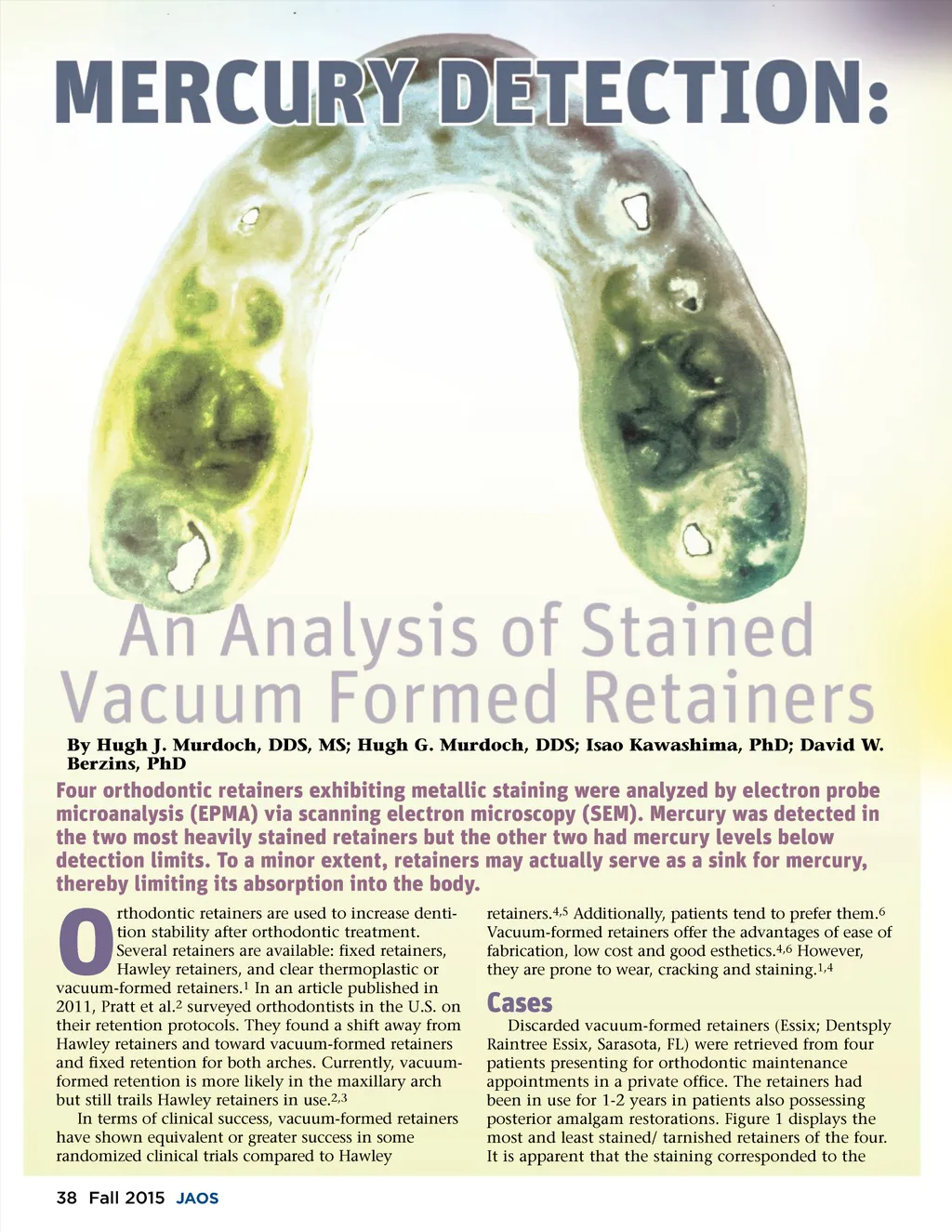
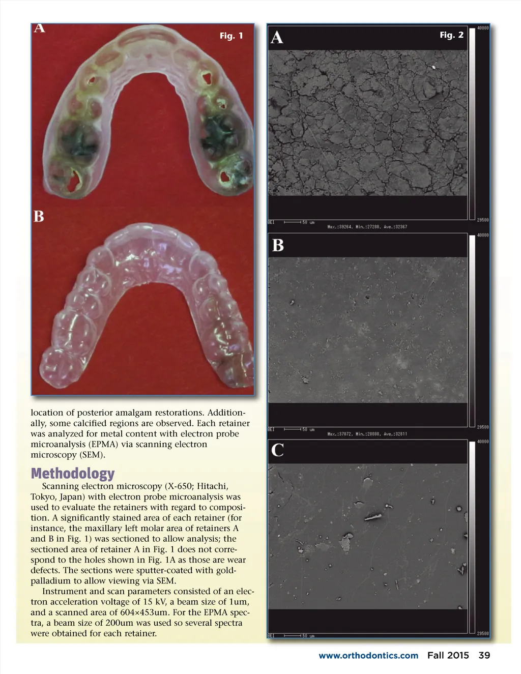
Fig. 1 Fig. 2 location of posterior amalgam restorations. Addition-ally, some calcified regions are observed. Each retainer was analyzed for metal content with electron probe microanalysis (EPMA) via scanning electron microscopy (SEM). Methodology Scanning electron microscopy (X-650; Hitachi, Tokyo, Japan) with electron probe microanalysis was used to evaluate the retainers with regard to composi-tion. A significantly stained area of each retainer (for instance, the maxillary left molar area of retainers A and B in Fig. 1) was sectioned to allow analysis; the sectioned area of retainer A in Fig. 1 does not corre-spond to the holes shown in Fig. 1A as those are wear defects. The sections were sputter-coated with gold-palladium to allow viewing via SEM. Instrument and scan parameters consisted of an elec-tron acceleration voltage of 15 kV, a beam size of 1um, and a scanned area of 604×453um. For the EPMA spec-tra, a beam size of 200um was used so several spectra were obtained for each retainer. www.orthodontics.com Fall 2015 39
Journal of the American Orthodontic Society Fall 2015: Page 39