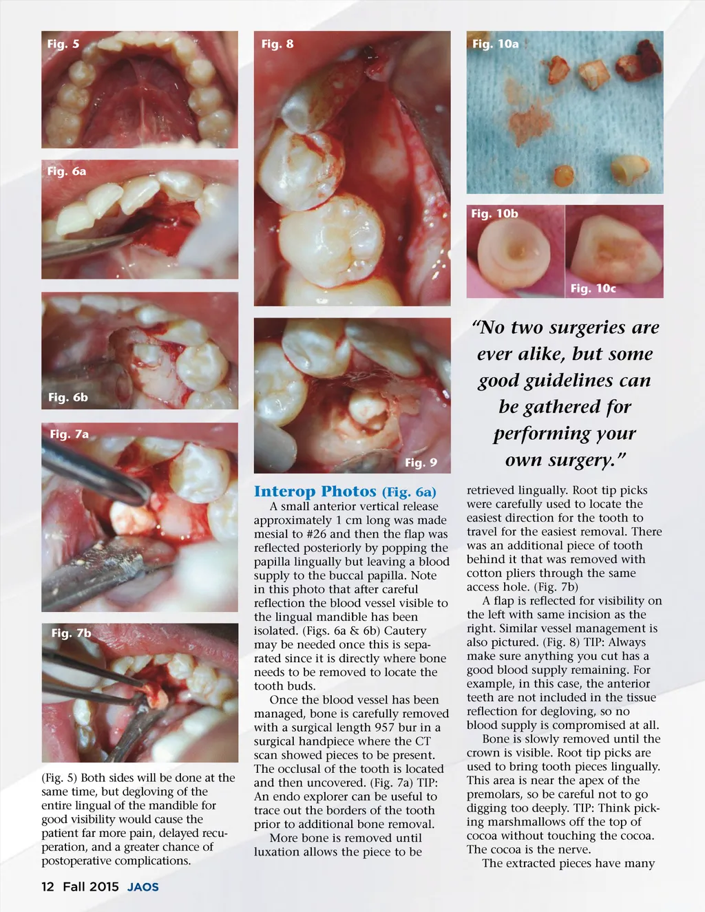
Fig. 11 Fig. 12b Fig. 12c Fig. 12e Fig. 12a Fig. 12d Fig. 12f different shapes and sizes as can be seen from the photos of the numer-ous pieces of supernumerary teeth that were removed. Resorption of some of the crowns that were touching adjacent teeth can be seen in Figs. 10b and c. knots are placed in the buccal inter-proximal space so that the patient does not contact them. (Fig. 11) 5-week Post-Op; Ready For Orthodontic Treatment Five weeks after the procedure, we can see that the patient is now ready for orthodontic treatment to start. (Figs. 12a-f) References 1. Schweiz Monatsschr Zahnmed. 2010;120 (11):987-93. 2. IJOPD Vol 12 244-254 July 2002 3. J Can Dent Assoc 1999; 65:612-6 4. J Am Dent Assoc. 1985 May;110 (5):721-3 Immediate Post-Op Photo Vicryl sutures are placed over the contacts to pull the tissue up. The www.orthodontics.com Fall 2015 13
Journal of the American Orthodontic Society Fall 2015: Page 13