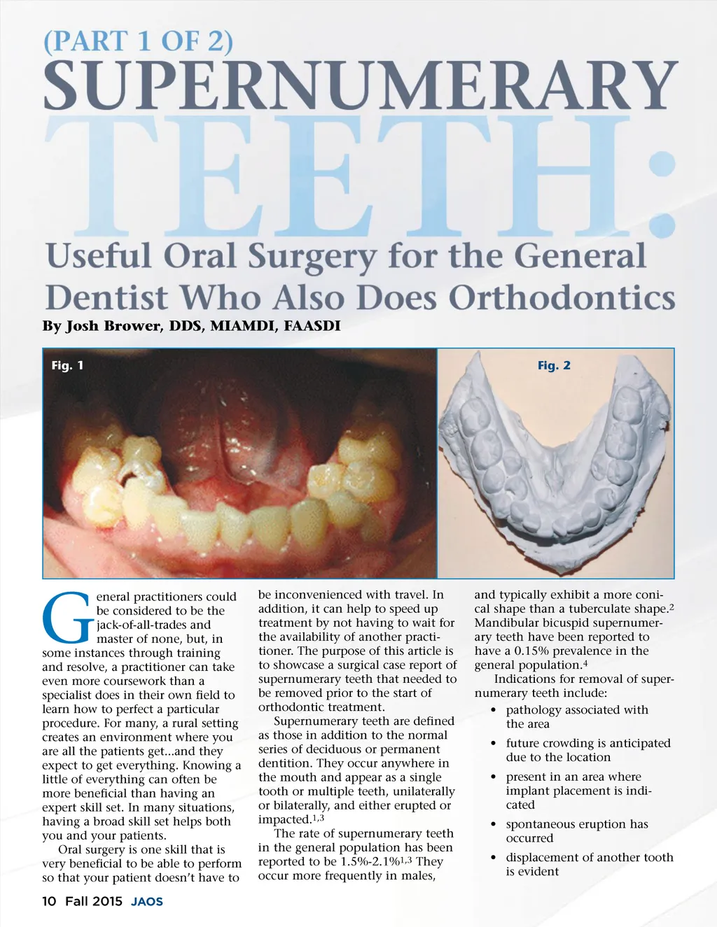
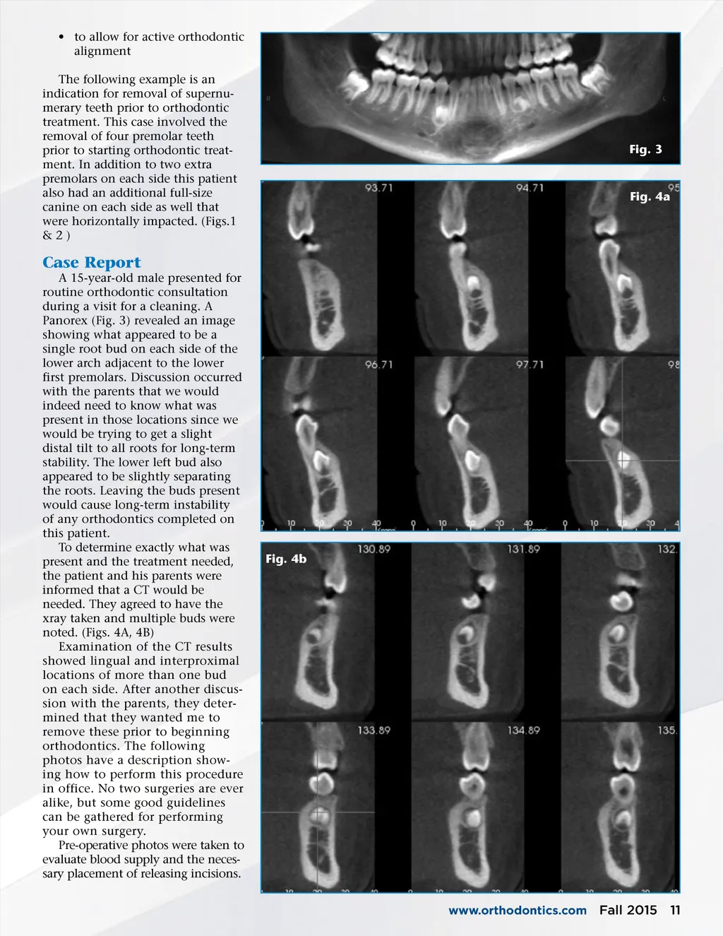
• to allow for active orthodontic alignment The following example is an indication for removal of supernu-merary teeth prior to orthodontic treatment. This case involved the removal of four premolar teeth prior to starting orthodontic treat-ment. In addition to two extra premolars on each side this patient also had an additional full-size canine on each side as well that were horizontally impacted. (Figs.1 & 2 ) Fig. 3 Fig. 4a Case Report A 15-year-old male presented for routine orthodontic consultation during a visit for a cleaning. A Panorex (Fig. 3) revealed an image showing what appeared to be a single root bud on each side of the lower arch adjacent to the lower first premolars. Discussion occurred with the parents that we would indeed need to know what was present in those locations since we would be trying to get a slight distal tilt to all roots for long-term stability. The lower left bud also appeared to be slightly separating the roots. Leaving the buds present would cause long-term instability of any orthodontics completed on this patient. To determine exactly what was present and the treatment needed, the patient and his parents were informed that a CT would be needed. They agreed to have the xray taken and multiple buds were noted. (Figs. 4A, 4B) Examination of the CT results showed lingual and interproximal locations of more than one bud on each side. After another discus-sion with the parents, they deter-mined that they wanted me to remove these prior to beginning orthodontics. The following photos have a description show-ing how to perform this procedure in office. No two surgeries are ever alike, but some good guidelines can be gathered for performing your own surgery. Pre-operative photos were taken to evaluate blood supply and the neces-sary placement of releasing incisions. Fig. 4b www.orthodontics.com Fall 2015 11
Journal of the American Orthodontic Society Fall 2015: Page 11