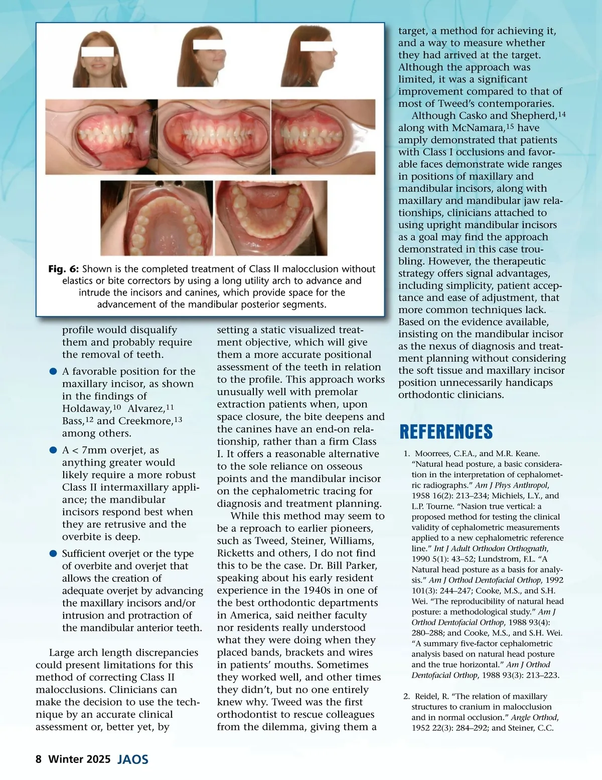
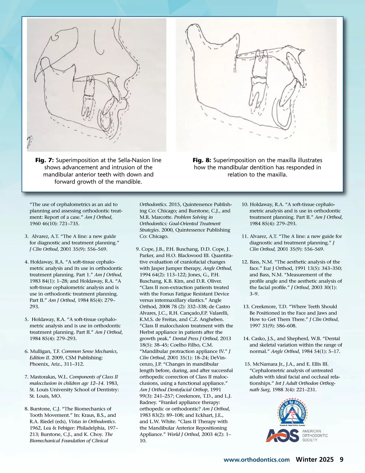
Fig. 7: Superimposition at the Sella-Nasion line shows advancement and intrusion of the mandibular anterior teeth with down and forward growth of the mandible. Fig. 8: Superimposition on the maxilla illustrates how the mandibular dentition has responded in relation to the maxilla. “The use of cephalometrics as an aid to planning and assessing orthodontic treat-ment: Report of a case.” Am J Orthod , 1960 46(10): 721–735. 3. Alvarez, A.T. “The A line: a new guide for diagnostic and treatment planning.” J Clin Orthod , 2001 35(9): 556–569. 4. Holdaway, R.A. “A soft-tissue cephalo-metric analysis and its use in orthodontic treatment planning. Part 1.” Am J Orthod , 1983 84(1): 1–28; and Holdaway, R.A. “A soft-tissue cephalometric analysis and is use in orthodontic treatment planning. Part II.” Am J Orthod , 1984 85(4): 279– 293. 5. Holdaway, R.A. “A soft-tissue cephalo-metric analysis and is use in orthodontic treatment planning. Part II.” Am J Orthod , 1984 85(4): 279–293. 6. Mulligan, T.F. Common Sense Mechanics, Edition II . 2009, CSM Publishing: Phoenix, Ariz., 311–312. 7. Mastorakas, W.L. Components of Class II malocclusion in children age 12–14 . 1983, St. Louis University School of Dentistry: St. Louis, MO. 8. Burstone, C.J. “The Biomechanics of Tooth Movement.” In: Kraus, B.S., and R.A. Riedel (eds), Vistas in Orthodontics . 1962, Lea & Febiger: Philadelphia, 197– 213; Burstone, C.J., and K. Choy. The Biomechanical Foundation of Clinical Orthodontics . 2015, Quintessence Publish-ing Co: Chicago; and Burstone, C.J., and M.R. Marcotte. Problem Solving in Orthodontics: Goal-Oriented Treatment Strategies . 2000, Quintessence Publishing Co: Chicago. 9. Cope, J.B., P.H. Buschang, D.D. Cope, J. Parker, and H.O. Blackwood III. Quantita-tive evaluation of craniofacial changes with Jasper Jumper therapy, Angle Orthod , 1994 64(2): 113–122; Jones, G., P.H. Buschang, K.B. Kim, and D.R. Oliver. “Class II non-extraction patients treated with the Forsus Fatigue Resistant Device versus intermaxillary elastics.” Angle Orthod, 2008 78 (2): 332–338; de Castro Alvares, J.C., R.H. Cançado,F.P. Valarelli, K.M.S. de Freitas, and C.Z. Angheben. “Class II malocclusion treatment with the Herbst appliance in patients after the growth peak.” Dental Press J Orthod , 2013 18(5): 38–45; Coelho Filho, C.M. “Mandibular protraction appliance IV.” J Clin Orthod , 2001 35(1): 18–24; DeVin-cenzo, J.P. “Changes in mandibular length before, during, and after successful orthopedic correction of Class II maloc-clusions, using a functional appliance.” Am J Orthod Dentofacial Orthop , 1991 99(3): 241–257; Creekmore, T.D., and L.J. Radney. “Frankel appliance therapy: orthopedic or orthodontic? Am J Orthod , 1983 83(2): 89–108; and Eckhart, J.E., and L.W. White. “Class II Therapy with the Mandibular Anterior Repositioning Appliance.” World J Orthod, 2003 4(2): 1– 10. 10. Holdaway, R.A. “A soft-tissue cephalo-metric analysis and is use in orthodontic treatment planning. Part II.” Am J Orthod , 1984 85(4): 279–293. 11. Alvarez, A.T. “The A line: a new guide for diagnostic and treatment planning.” J Clin Orthod, 2001 35(9): 556–569. 12. Bass, N.M. “The aesthetic analysis of the face.” Eur J Orthod, 1991 13(5): 343–350; and Bass, N.M. “Measurement of the profile angle and the aesthetic analysis of the facial profile.” J Orthod , 2003 30(1): 3–9. 13. Creekmore, T.D. “Where Teeth Should Be Positioned in the Face and Jaws and How to Get Them There.” J Clin Orthod , 1997 31(9): 586–608. 14. Casko, J.S., and Shepherd, W.B. “Dental and skeletal variation within the range of normal.” Angle Orthod , 1984 54(1): 5–17. 15. McNamara Jr., J.A., and E. Ellis III. “Cephalometric analysis of untreated adults with ideal facial and occlusal rela-tionships.” Int J Adult Orthodon Orthog-nath Surg , 1988 3(4): 221–231. www.orthodontics.com Winter 2025 9
Journal of the American Orthodontic Society Winter 2025: Page 9