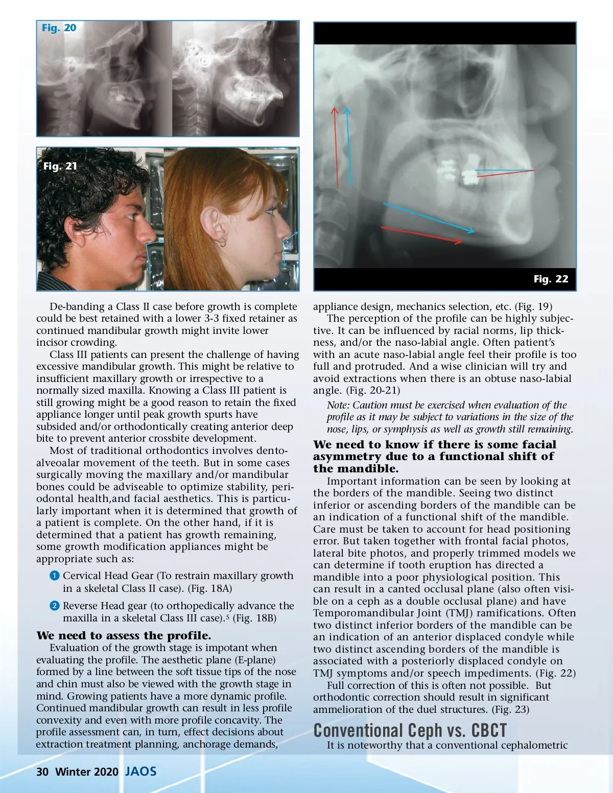
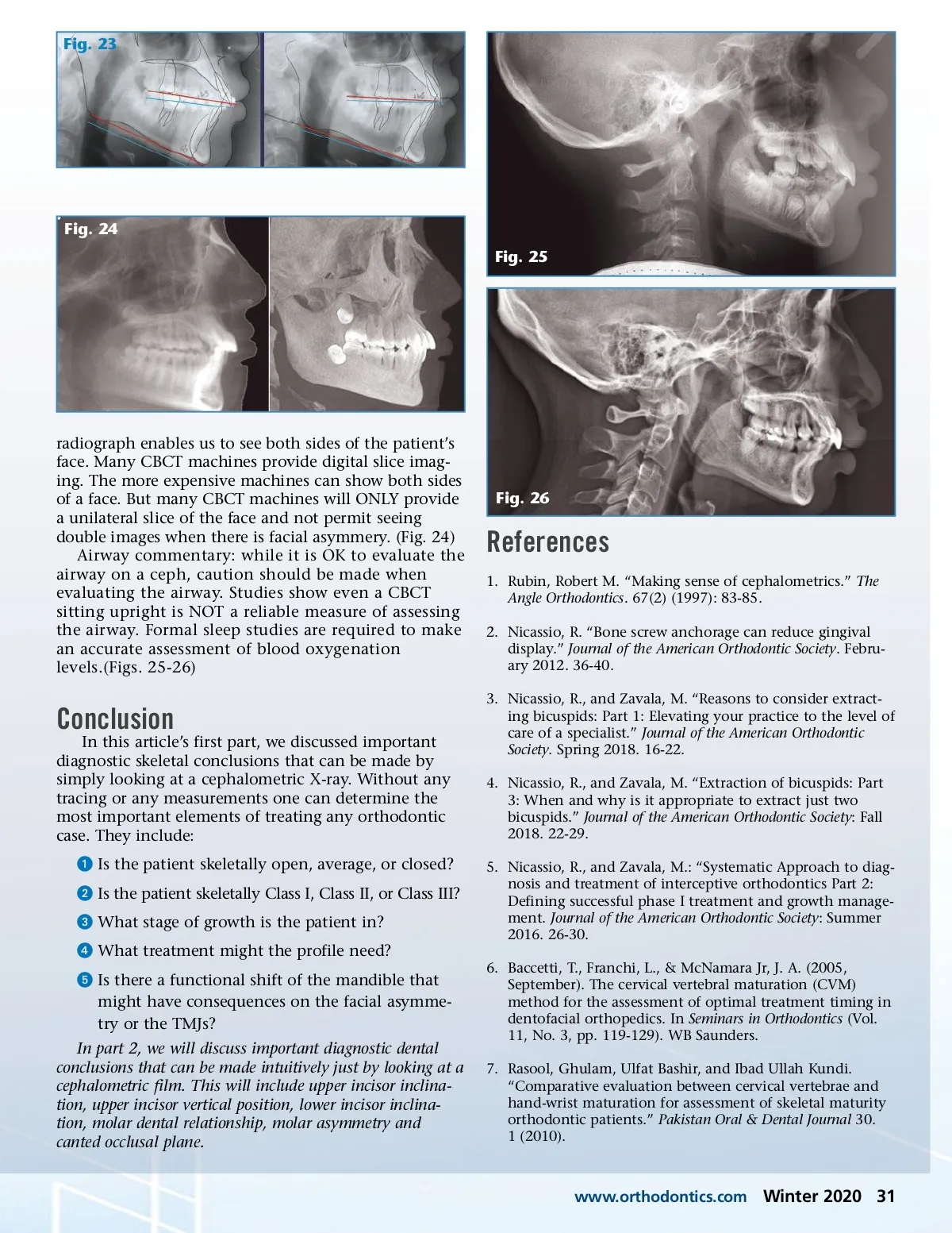
Fig. 23 Fig. 24 Fig. 25 radiograph enables us to see both sides of the patient’s face. Many CBCT machines provide digital slice imag-ing. The more expensive machines can show both sides of a face. But many CBCT machines will ONLY provide a unilateral slice of the face and not permit seeing double images when there is facial asymmery. (Fig. 24) Airway commentary: while it is OK to evaluate the airway on a ceph, caution should be made when evaluating the airway. Studies show even a CBCT sitting upright is NOT a reliable measure of assessing the airway. Formal sleep studies are required to make an accurate assessment of blood oxygenation levels.(Figs. 25-26) Fig. 26 References 1. Rubin, Robert M. “Making sense of cephalometrics.” The Angle Orthodontics. 67(2) (1997): 83-85. 2. Nicassio, R. “Bone screw anchorage can reduce gingival display.” Journal of the American Orthodontic Society . Febru-ary 2012. 36-40. 3. Nicassio, R., and Zavala, M. “Reasons to consider extract-ing bicuspids: Part 1: Elevating your practice to the level of care of a specialist.” Journal of the American Orthodontic Society . Spring 2018. 16-22. 4. Nicassio, R., and Zavala, M. “Extraction of bicuspids: Part 3: When and why is it appropriate to extract just two bicuspids.” Journal of the American Orthodontic Society : Fall 2018. 22-29. 5. Nicassio, R., and Zavala, M.: “Systematic Approach to diag-nosis and treatment of interceptive orthodontics Part 2: Defining successful phase I treatment and growth manage-ment. Journal of the American Orthodontic Society : Summer 2016. 26-30. 6. Baccetti, T., Franchi, L., & McNamara Jr, J. A. (2005, September). The cervical vertebral maturation (CVM) method for the assessment of optimal treatment timing in dentofacial orthopedics. In Seminars in Orthodontics (Vol. 11, No. 3, pp. 119-129). WB Saunders. 7. Rasool, Ghulam, Ulfat Bashir, and Ibad Ullah Kundi. “Comparative evaluation between cervical vertebrae and hand-wrist maturation for assessment of skeletal maturity orthodontic patients.” Pakistan Oral & Dental Journal 30. 1 (2010). Conclusion In this article’s first part, we discussed important diagnostic skeletal conclusions that can be made by simply looking at a cephalometric X-ray. Without any tracing or any measurements one can determine the most important elements of treating any orthodontic case. They include: ᕡ Is the patient skeletally open, average, or closed? ᕢ Is the patient skeletally Class I, Class II, or Class III? ᕣ What stage of growth is the patient in? ᕤ What treatment might the profile need? ᕥ Is there a functional shift of the mandible that might have consequences on the facial asymme-try or the TMJs? In part 2, we will discuss important diagnostic dental conclusions that can be made intuitively just by looking at a cephalometric film. This will include upper incisor inclina-tion, upper incisor vertical position, lower incisor inclina-tion, molar dental relationship, molar asymmetry and canted occlusal plane. www.orthodontics.com Winter 2020 31
Journal of the American Orthodontic Society Winter 2020: Page 31