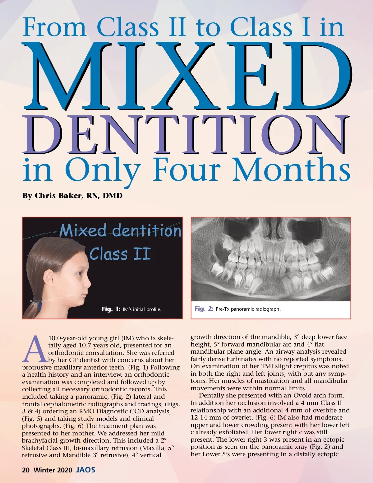
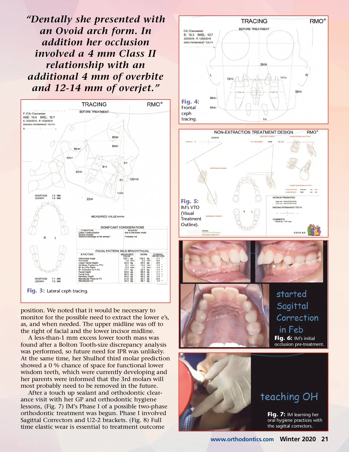
MIXED DENTITION in Only Four Months By Chris Baker, RN, DMD Fig. 1: IM’s initial profile. From Class II to Class I in Fig. 2: Pre-Tx panoramic radiograph. A 10.0-year-old young girl (IM) who is skele-tally aged 10.7 years old, presented for an orthodontic consultation. She was referred by her GP dentist with concerns about her protrusive maxillary anterior teeth. (Fig. 1) Following a health history and an interview, an orthodontic examination was completed and followed up by collecting all necessary orthodontic records. This included taking a panoramic, (Fig. 2) lateral and frontal cephalometric radiographs and tracings, (Figs. 3 & 4) ordering an RMO Diagnostic CCD analysis, (Fig. 5) and taking study models and clinical photographs. (Fig. 6) The treatment plan was presented to her mother. We addressed her mild brachyfacial growth direction. This included a 2° Skeletal Class III, bi-maxillary retrusion (Maxilla, 5° retrusive and Mandible 3° retrusive), 4° vertical growth direction of the mandible, 3° deep lower face height, 5° forward mandibular arc and 4° flat mandibular plane angle. An airway analysis revealed fairly dense turbinates with no reported symptoms. On examination of her TMJ slight crepitus was noted in both the right and left joints, with out any symp-toms. Her muscles of mastication and all mandibular movements were within normal limits. Dentally she presented with an Ovoid arch form. In addition her occlusion involved a 4 mm Class II relationship with an additional 4 mm of overbite and 12-14 mm of overjet. (Fig. 6) IM also had moderate upper and lower crowding present with her lower left c already exfoliated. Her lower right c was still present. The lower right 3 was present in an ectopic position as seen on the panoramic xray (Fig. 2) and her Lower 5’s were presenting in a distally ectopic 20 Winter 2020 JAOS
Journal of the American Orthodontic Society Winter 2020: Page 20