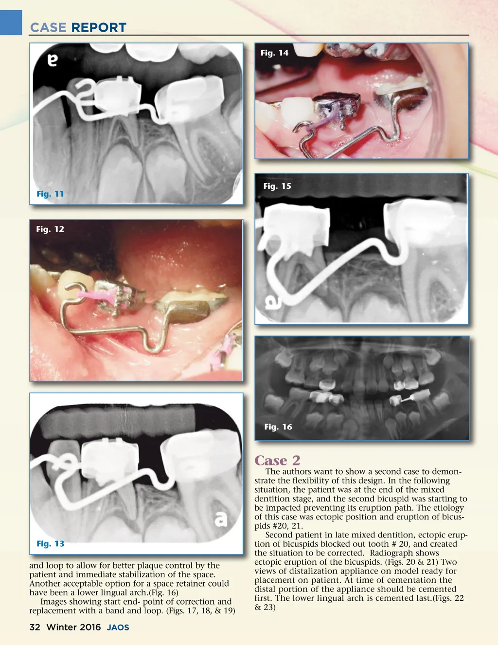
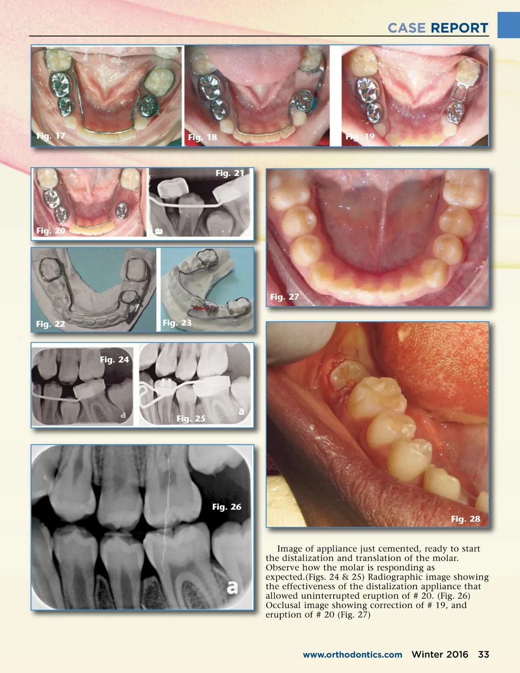
CASE REPORT Fig. 17 Fig. 18 Fig. 19 Fig. 21 Fig. 20 Fig. 27 Fig. 22 Fig. 23 Fig. 24 Fig. 25 Fig. 26 Fig. 28 Image of appliance just cemented, ready to start the distalization and translation of the molar. Observe how the molar is responding as expected.(Figs. 24 & 25) Radiographic image showing the effectiveness of the distalization appliance that allowed uninterrupted eruption of # 20. (Fig. 26) Occlusal image showing correction of # 19, and eruption of # 20 (Fig. 27) www.orthodontics.com Winter 2016 33
Journal of the American Orthodontic Society Winter 2016: Page 33