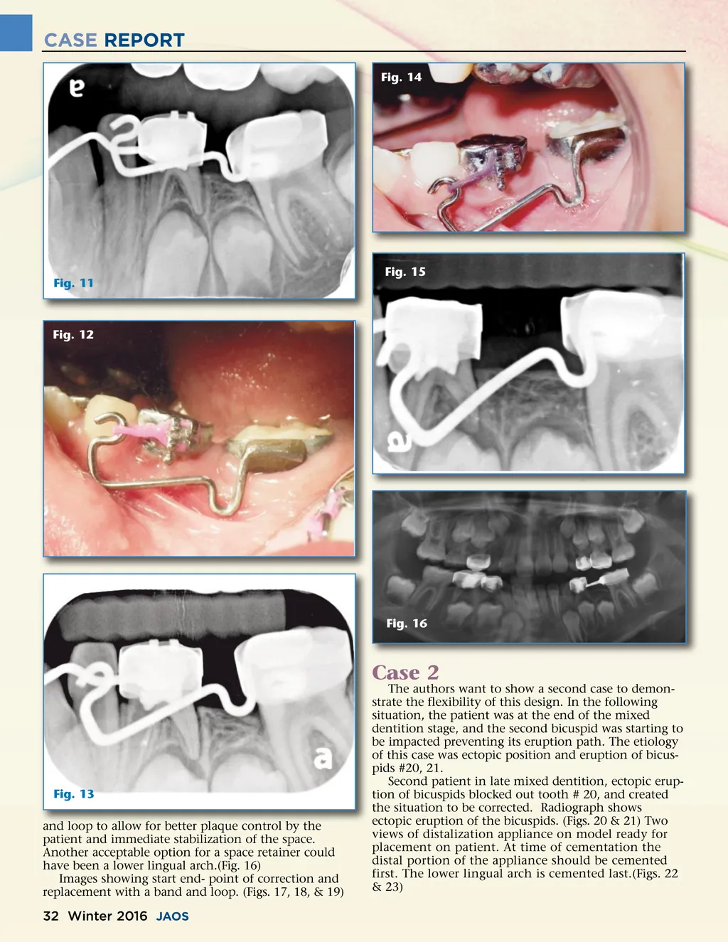
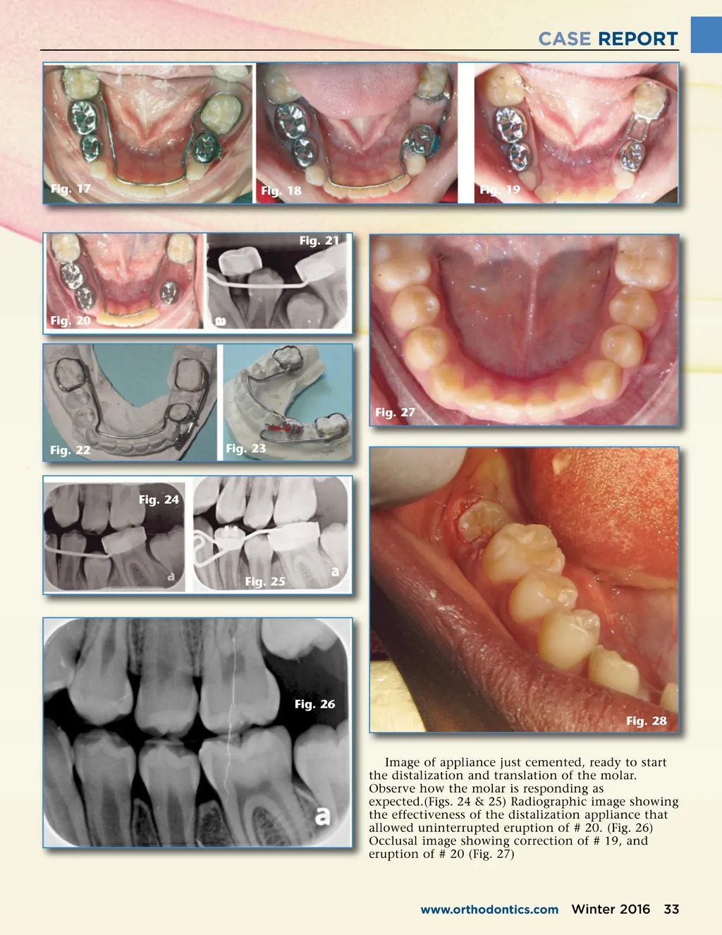
CASE REPORT Fig. 14 Fig. 11 Fig. 15 Fig. 12 Fig. 16 Fig. 13 and loop to allow for better plaque control by the patient and immediate stabilization of the space. Another acceptable option for a space retainer could have been a lower lingual arch.(Fig. 16) Images showing start end-point of correction and replacement with a band and loop. (Figs. 17, 18, & 19) The authors want to show a second case to demon-strate the flexibility of this design. In the following situation, the patient was at the end of the mixed dentition stage, and the second bicuspid was starting to be impacted preventing its eruption path. The etiology of this case was ectopic position and eruption of bicus-pids #20, 21. Second patient in late mixed dentition, ectopic erup-tion of bicuspids blocked out tooth # 20, and created the situation to be corrected. Radiograph shows ectopic eruption of the bicuspids. (Figs. 20 & 21) Two views of distalization appliance on model ready for placement on patient. At time of cementation the distal portion of the appliance should be cemented first. The lower lingual arch is cemented last.(Figs. 22 & 23) Case 2 32 Winter 2016 JAOS
Journal of the American Orthodontic Society Winter 2016: Page 32