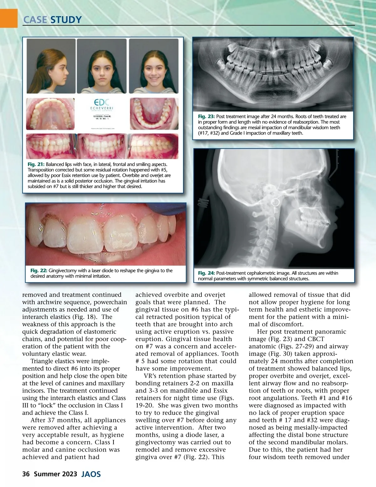
CASE STUDY Fig. 23: Post treatment image after 24 months. Roots of teeth treated are in proper form and length with no evidence of reabsorption. The most outstanding findings are mesial impaction of mandibular wisdom teeth (#17, #32) and Grade I impaction of maxillary teeth. Fig. 21: Balanced lips with face, in lateral, frontal and smiling aspects. Transposition corrected but some residual rotation happened with #5, allowed by poor Essix retention use by patient. Overbite and overjet are maintained as is a solid posterior occlusion. The gingival irritation has subsided on #7 but is still thicker and higher that desired. Fig. 22: Gingivectomy with a laser diode to reshape the gingiva to the desired anatomy with minimal irritation. Fig. 24: Post-treatment cephalometric image. All structures are within normal parameters with symmetric balanced structures. removed and treatment continued with archwire sequence, powerchain adjustments as needed and use of interarch elastics (Fig. 18). The weakness of this approach is the quick degradation of elastomeric chains, and potential for poor coop-eration of the patient with the voluntary elastic wear. Triangle elastics were imple-mented to direct #6 into its proper position and help close the open bite at the level of canines and maxillary incisors. The treatment continued using the interarch elastics and Class III to “lock” the occlusion in Class I and achieve the Class I. After 37 months, all appliances were removed after achieving a very acceptable result, as hygiene had become a concern. Class I molar and canine occlusion was achieved and patient had achieved overbite and overjet goals that were planned. The gingival tissue on #6 has the typi-cal retracted position typical of teeth that are brought into arch using active eruption vs. passive eruption. Gingival tissue health on #7 was a concern and acceler-ated removal of appliances. Tooth # 5 had some rotation that could have some improvement. VR’s retention phase started by bonding retainers 2-2 on maxilla and 3-3 on mandible and Essix retainers for night time use (Figs. 19-20. She was given two months to try to reduce the gingival swelling over #7 before doing any active intervention. After two months, using a diode laser, a gingivectomy was carried out to remodel and remove excessive gingiva over #7 (Fig. 22). This allowed removal of tissue that did not allow proper hygiene for long term health and esthetic improve-ment for the patient with a mini-mal of discomfort. Her post treatment panoramic image (Fig. 23) and CBCT anatomic (Figs. 27-29) and airway image (Fig. 30) taken approxi-mately 24 months after completion of treatment showed balanced lips, proper overbite and overjet, excel-lent airway flow and no reabsorp-tion of teeth or roots, with proper root angulations. Teeth #1 and #16 were diagnosed as impacted with no lack of proper eruption space and teeth # 17 and #32 were diag-nosed as being mesially-impacted affecting the distal bone structure of the second mandibular molars. Due to this, the patient had her four wisdom teeth removed under 36 Summer 2023 JAOS
Journal of the American Orthodontic Society Summer 2023: Page 36