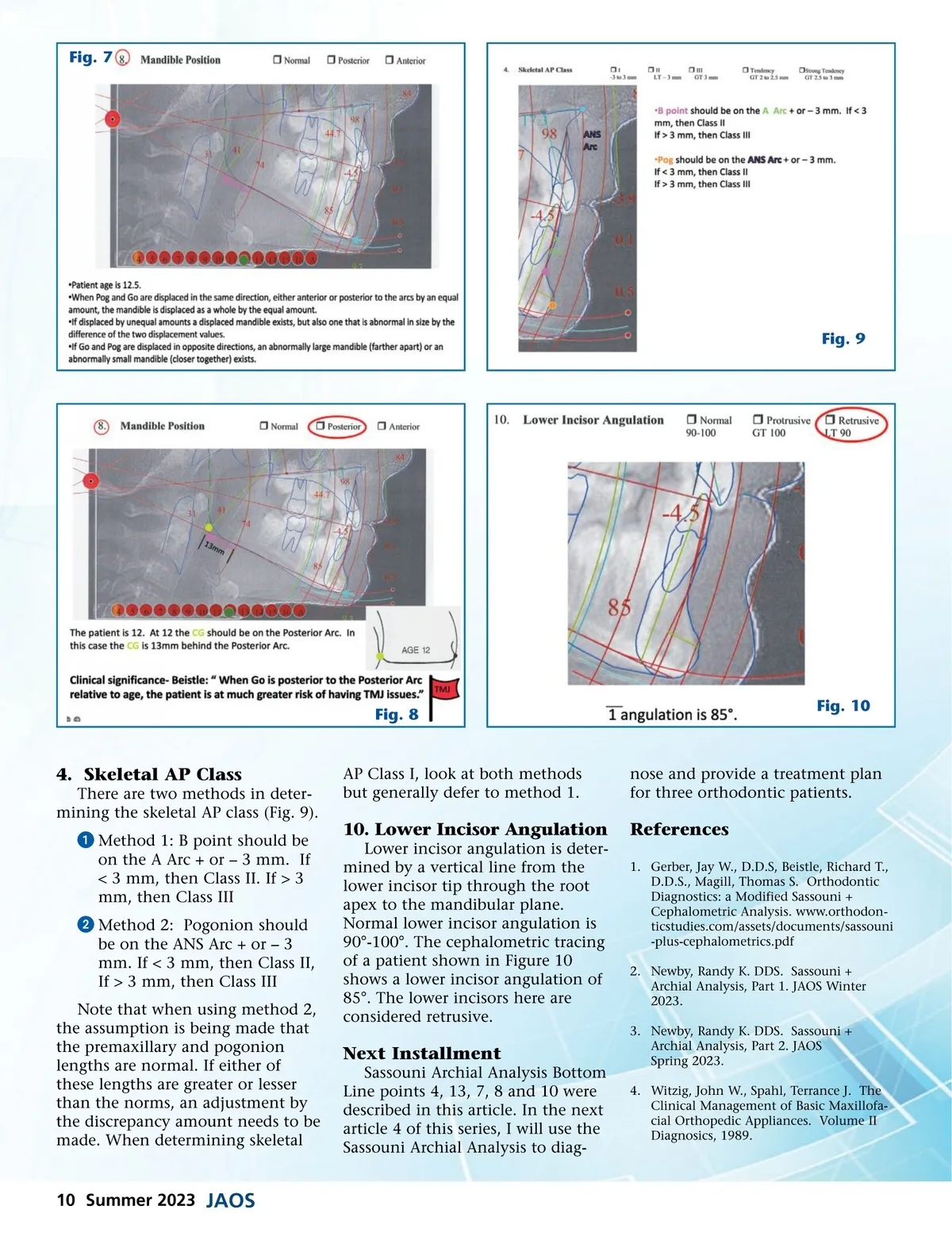
Fig. 7 Fig. 9 Fig. 8 Fig. 10 4. Skeletal AP Class There are two methods in deter-mining the skeletal AP class (Fig. 9). AP Class I, look at both methods but generally defer to method 1. nose and provide a treatment plan for three orthodontic patients. ᕡ Method 1: B point should be on the A Arc + or – 3 mm. If < 3 mm, then Class II. If > 3 mm, then Class III 10. Lower Incisor Angulation Lower incisor angulation is deter-mined by a vertical line from the lower incisor tip through the root apex to the mandibular plane. Normal lower incisor angulation is 90°-100°. The cephalometric tracing of a patient shown in Figure 10 shows a lower incisor angulation of 85°. The lower incisors here are considered retrusive. References 1. Gerber, Jay W., D.D.S, Beistle, Richard T., D.D.S., Magill, Thomas S. Orthodontic Diagnostics: a Modified Sassouni + Cephalometric Analysis. www.orthodon-ticstudies.com/assets/documents/sassouni -plus-cephalometrics.pdf 2. Newby, Randy K. DDS. Sassouni + Archial Analysis, Part 1. JAOS Winter 2023. 3. Newby, Randy K. DDS. Sassouni + Archial Analysis, Part 2. JAOS Spring 2023. 4. Witzig, John W., Spahl, Terrance J. The Clinical Management of Basic Maxillofa-cial Orthopedic Appliances. Volume II Diagnosics, 1989. ᕢ Method 2: Pogonion should be on the ANS Arc + or – 3 mm. If < 3 mm, then Class II, If > 3 mm, then Class III Note that when using method 2, the assumption is being made that the premaxillary and pogonion lengths are normal. If either of these lengths are greater or lesser than the norms, an adjustment by the discrepancy amount needs to be made. When determining skeletal Next Installment Sassouni Archial Analysis Bottom Line points 4, 13, 7, 8 and 10 were described in this article. In the next article 4 of this series, I will use the Sassouni Archial Analysis to diag-10 Summer 2023 JAOS
Journal of the American Orthodontic Society Summer 2023: Page 10