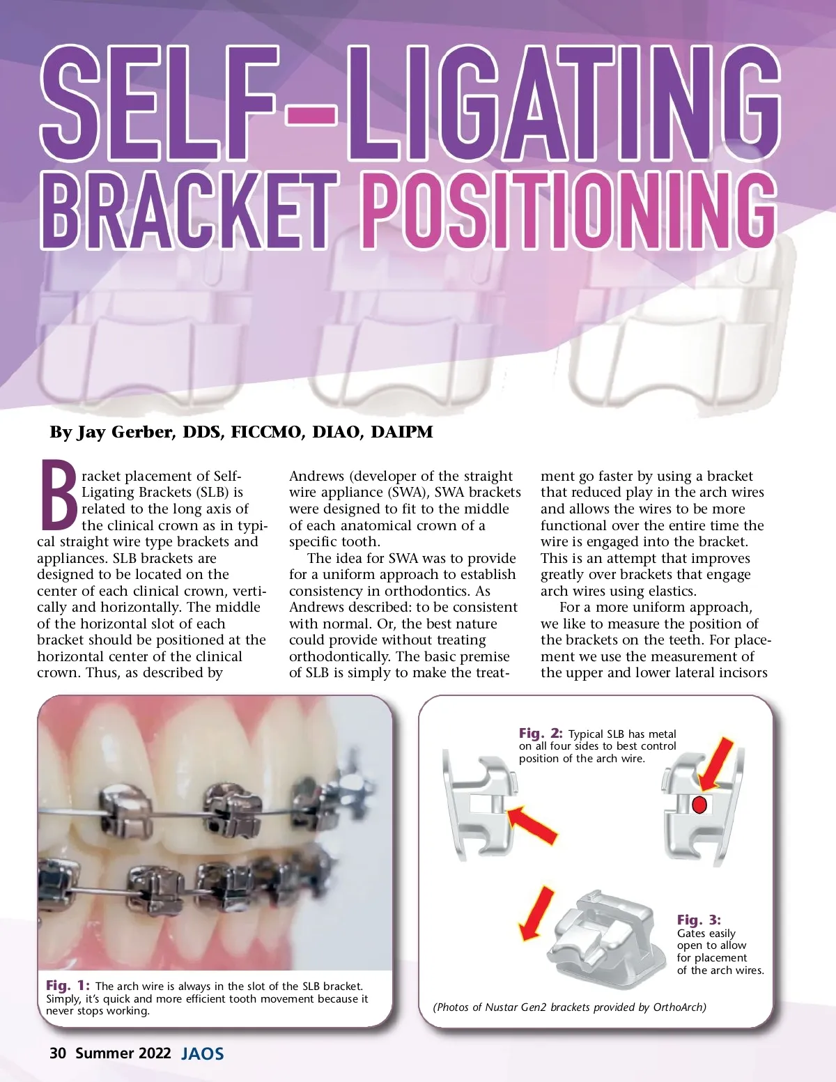
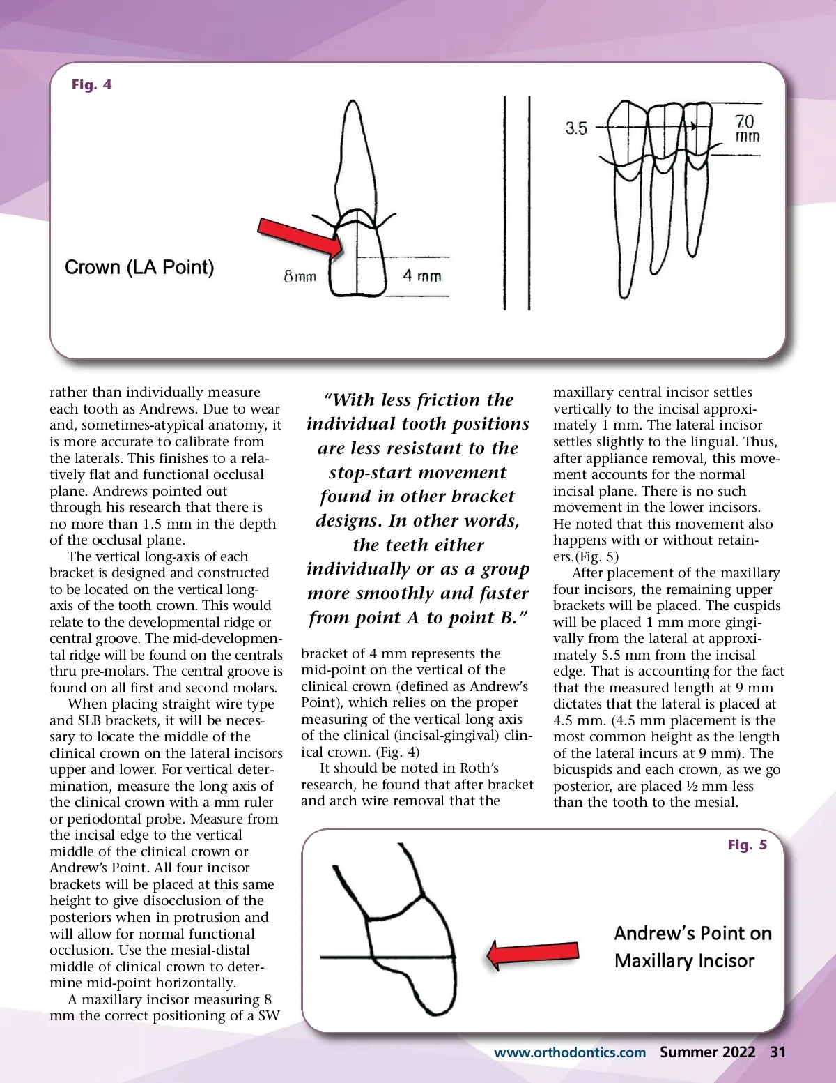
Fig. 4 rather than individually measure each tooth as Andrews. Due to wear and, sometimes-atypical anatomy, it is more accurate to calibrate from the laterals. This finishes to a rela-tively flat and functional occlusal plane. Andrews pointed out through his research that there is no more than 1.5 mm in the depth of the occlusal plane. The vertical long-axis of each bracket is designed and constructed to be located on the vertical long-axis of the tooth crown. This would relate to the developmental ridge or central groove. The mid-developmen-tal ridge will be found on the centrals thru pre-molars. The central groove is found on all first and second molars. When placing straight wire type and SLB brackets, it will be neces-sary to locate the middle of the clinical crown on the lateral incisors upper and lower. For vertical deter-mination, measure the long axis of the clinical crown with a mm ruler or periodontal probe. Measure from the incisal edge to the vertical middle of the clinical crown or Andrew’s Point. All four incisor brackets will be placed at this same height to give disocclusion of the posteriors when in protrusion and will allow for normal functional occlusion. Use the mesial-distal middle of clinical crown to deter-mine mid-point horizontally. A maxillary incisor measuring 8 mm the correct positioning of a SW “With less friction the individual tooth positions are less resistant to the stop-start movement found in other bracket designs. In other words, the teeth either individually or as a group more smoothly and faster from point A to point B.” bracket of 4 mm represents the mid-point on the vertical of the clinical crown (defined as Andrew’s Point), which relies on the proper measuring of the vertical long axis of the clinical (incisal-gingival) clin-ical crown. (Fig. 4) It should be noted in Roth’s research, he found that after bracket and arch wire removal that the maxillary central incisor settles vertically to the incisal approxi-mately 1 mm. The lateral incisor settles slightly to the lingual. Thus, after appliance removal, this move-ment accounts for the normal incisal plane. There is no such movement in the lower incisors. He noted that this movement also happens with or without retain-ers.(Fig. 5) After placement of the maxillary four incisors, the remaining upper brackets will be placed. The cuspids will be placed 1 mm more gingi-vally from the lateral at approxi-mately 5.5 mm from the incisal edge. That is accounting for the fact that the measured length at 9 mm dictates that the lateral is placed at 4.5 mm. (4.5 mm placement is the most common height as the length of the lateral incurs at 9 mm). The bicuspids and each crown, as we go posterior, are placed ½ mm less than the tooth to the mesial. Fig. 5 www.orthodontics.com Summer 2022 31
Journal of the American Orthodontic Society Summer 2022: Page 31