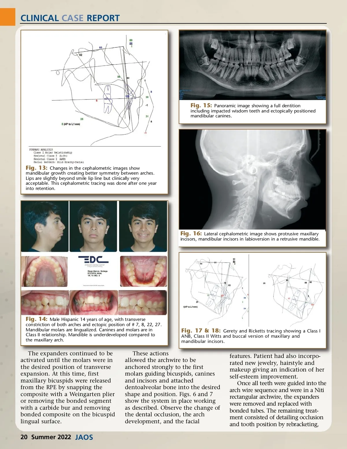
CLINICAL CASE REPORT Fig. 15: Panoramic image showing a full dentition including impacted wisdom teeth and ectopically positioned mandibular canines. Fig. 13: Changes in the cephalometric images show mandibular growth creating better symmetry between arches. Lips are slightly beyond smile lip line but clinically very acceptable. This cephalometric tracing was done after one year into retention. Fig. 16: Lateral cephalometric image shows protrusive maxillary incisors, mandibular incisors in labioversion in a retrusive mandible. Fig. 14: Male Hispanic 14 years of age, with transverse constriction of both arches and ectopic position of # 7, 8, 22, 27. Mandibular molars are lingualized. Canines and molars are in Class II relationship. Mandible is underdeveloped compared to the maxillary arch. Fig. 17 & 18: Gerety and Ricketts tracing showing a Class I ANB, Class II Witts and buccal version of maxillary and mandibular incisors. The expanders continued to be activated until the molars were in the desired position of transverse expansion. At this time, first maxillary bicuspids were released from the RPE by snapping the composite with a Weingarten plier or removing the bonded segment with a carbide bur and removing bonded composite on the bicuspid lingual surface. These actions allowed the archwire to be anchored strongly to the first molars guiding bicuspids, canines and incisors and attached dentoalveolar bone into the desired shape and position. Figs. 6 and 7 show the system in place working as described. Observe the change of the dental occlusion, the arch development, and the facial features. Patient had also incorpo-rated new jewelry, hairstyle and makeup giving an indication of her self-esteem improvement. Once all teeth were guided into the arch wire sequence and were in a Niti rectangular archwire, the expanders were removed and replaced with bonded tubes. The remaining treat-ment consisted of detailing occlusion and tooth position by rebracketing, 20 Summer 2022 JAOS
Journal of the American Orthodontic Society Summer 2022: Page 20