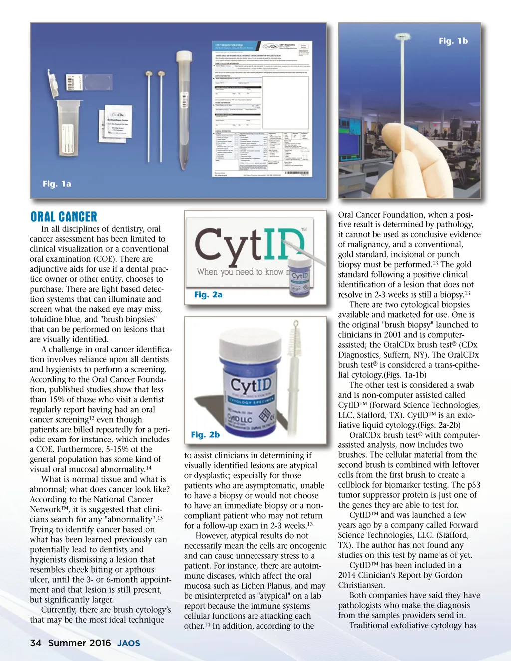
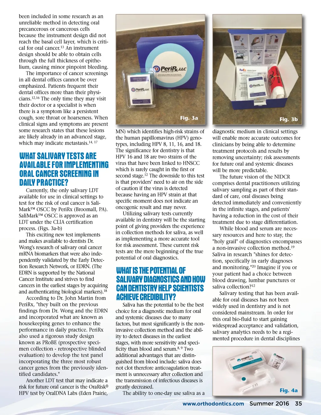
Fig. 1b Fig. 1a ORAL CANCER In all disciplines of dentistry, oral cancer assessment has been limited to clinical visualization or a conventional oral examination (COE). There are adjunctive aids for use if a dental prac-tice owner or other entity, chooses to purchase. There are light based detec-tion systems that can illuminate and screen what the naked eye may miss, toluidine blue, and "brush biopsies" that can be performed on lesions that are visually identified. A challenge in oral cancer identifica-tion involves reliance upon all dentists and hygienists to perform a screening. According to the Oral Cancer Founda-tion, published studies show that less than 15% of those who visit a dentist regularly report having had an oral cancer screening 13 even though patients are billed repeatedly for a peri-odic exam for instance, which includes a COE. Furthermore, 5-15% of the general population has some kind of visual oral mucosal abnormality. 14 What is normal tissue and what is abnormal; what does cancer look like? According to the National Cancer Network™, it is suggested that clini-cians search for any "abnormality". 15 Trying to identify cancer based on what has been learned previously can potentially lead to dentists and hygienists dismissing a lesion that resembles cheek biting or apthous ulcer, until the 3-or 6-month appoint-ment and that lesion is still present, but significantly larger. Currently, there are brush cytology’s that may be the most ideal technique Fig. 2a Fig. 2b to assist clinicians in determining if visually identified lesions are atypical or dysplastic; especially for those patients who are asymptomatic, unable to have a biopsy or would not choose to have an immediate biopsy or a non-compliant patient who may not return for a follow-up exam in 2-3 weeks. 13 However, atypical results do not necessarily mean the cells are oncogenic and can cause unnecessary stress to a patient. For instance, there are autoim-mune diseases, which affect the oral mucosa such as Lichen Planus, and may be misinterpreted as "atypical" on a lab report because the immune systems cellular functions are attacking each other. 14 In addition, according to the Oral Cancer Foundation, when a posi-tive result is determined by pathology, it cannot be used as conclusive evidence of malignancy, and a conventional, gold standard, incisional or punch biopsy must be performed. 13 The gold standard following a positive clinical identification of a lesion that does not resolve in 2-3 weeks is still a biopsy. 13 There are two cytological biopsies available and marketed for use. One is the original "brush biopsy" launched to clinicians in 2001 and is computer-assisted; the OralCDx brush test ® (CDx Diagnostics, Suffern, NY). The OralCDx brush test ® is considered a trans-epithe-lial cytology.(Figs. 1a-1b) The other test is considered a swab and is non-computer assisted called CytID™ (Forward Science Technologies, LLC. Stafford, TX). CytID™ is an exfo-liative liquid cytology.(Figs. 2a-2b) OralCDx brush test ® with computer-assisted analysis, now includes two brushes. The cellular material from the second brush is combined with leftover cells from the first brush to create a cellblock for biomarker testing. The p53 tumor suppressor protein is just one of the genes they are able to test for. CytID™ and was launched a few years ago by a company called Forward Science Technologies, LLC. (Stafford, TX). The author has not found any studies on this test by name as of yet. CytID™ has been included in a 2014 Clinician’s Report by Gordon Christiansen. Both companies have said they have pathologists who make the diagnosis from the samples providers send in. Traditional exfoliative cytology has 34 Summer 2016 JAOS
Journal of the American Orthodontic Society Summer 2016 / Buyer's Guide: Page 34