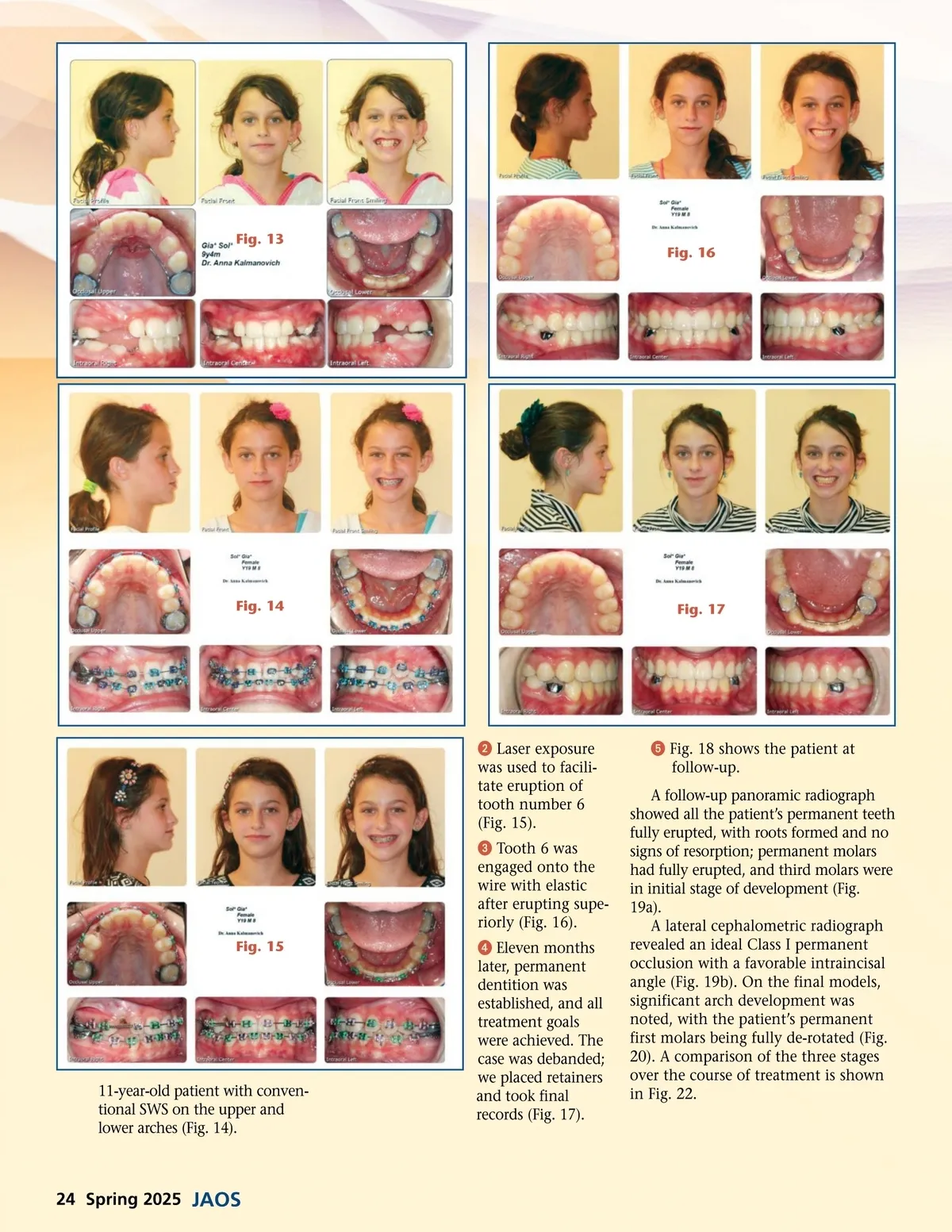
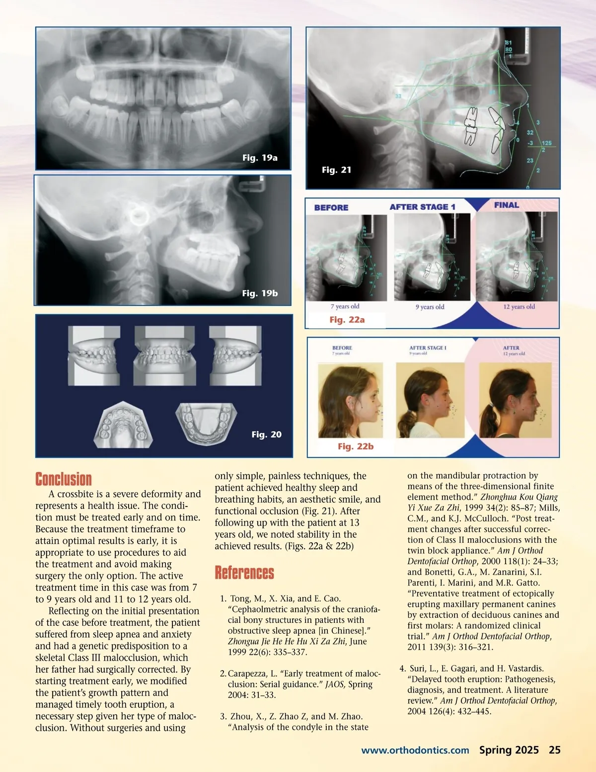
Fig. 13 Fig. 16 Fig. 14 Fig. 17 ᕢ Laser exposure was used to facili-tate eruption of tooth number 6 (Fig. 15). ᕣ Tooth 6 was engaged onto the wire with elastic after erupting supe-riorly (Fig. 16). Fig. 15 ᕥ Fig. 18 shows the patient at follow-up. A follow-up panoramic radiograph showed all the patient’s permanent teeth fully erupted, with roots formed and no signs of resorption; permanent molars had fully erupted, and third molars were in initial stage of development (Fig. 19a). A lateral cephalometric radiograph revealed an ideal Class I permanent occlusion with a favorable intraincisal angle (Fig. 19b). On the final models, significant arch development was noted, with the patient’s permanent first molars being fully de-rotated (Fig. 20). A comparison of the three stages over the course of treatment is shown in Fig. 22. 11-year-old patient with conven-tional SWS on the upper and lower arches (Fig. 14). ᕤ Eleven months later, permanent dentition was established, and all treatment goals were achieved. The case was debanded; we placed retainers and took final records (Fig. 17). 24 Spring 2025 JAOS
Journal of the American Orthodontic Society Spring 2025: Page 24