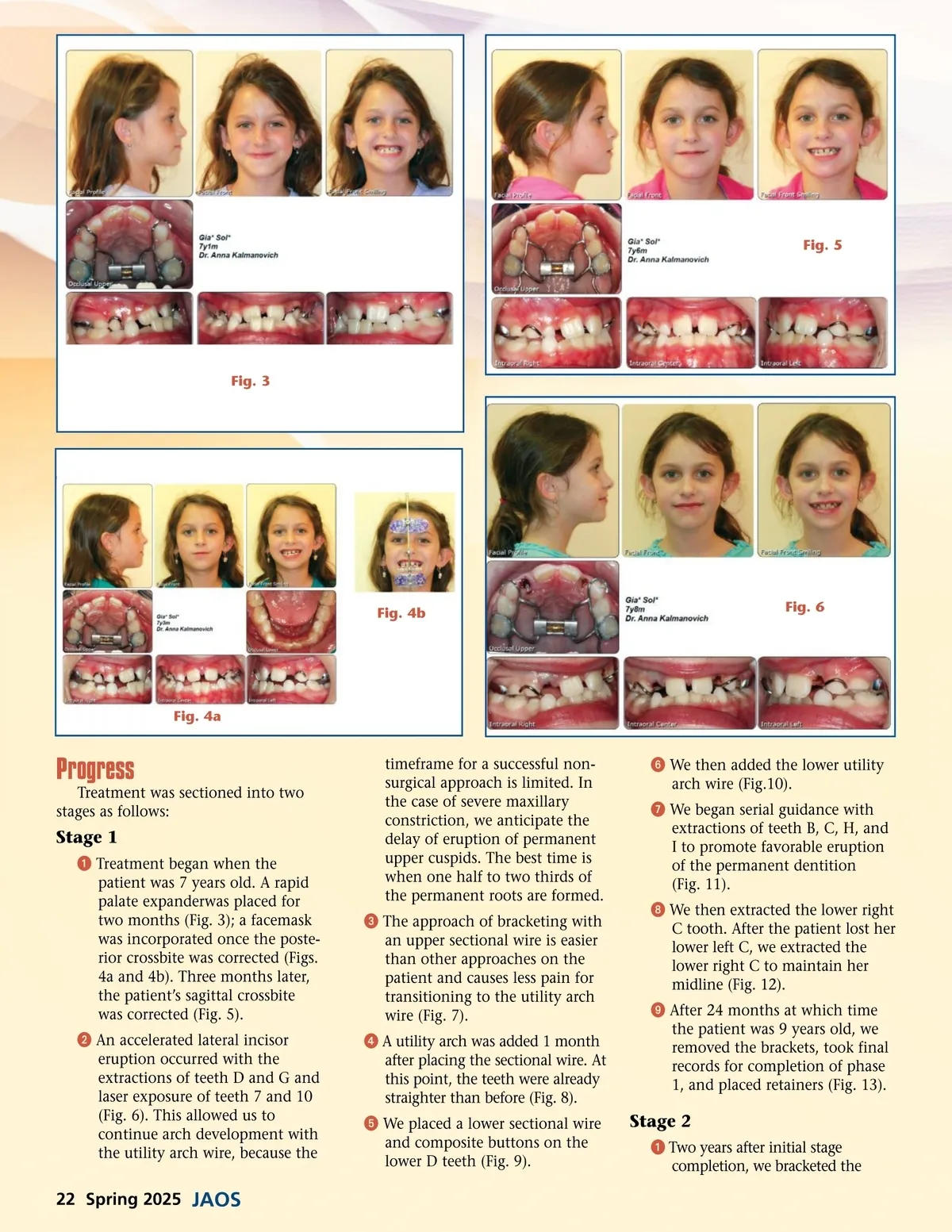
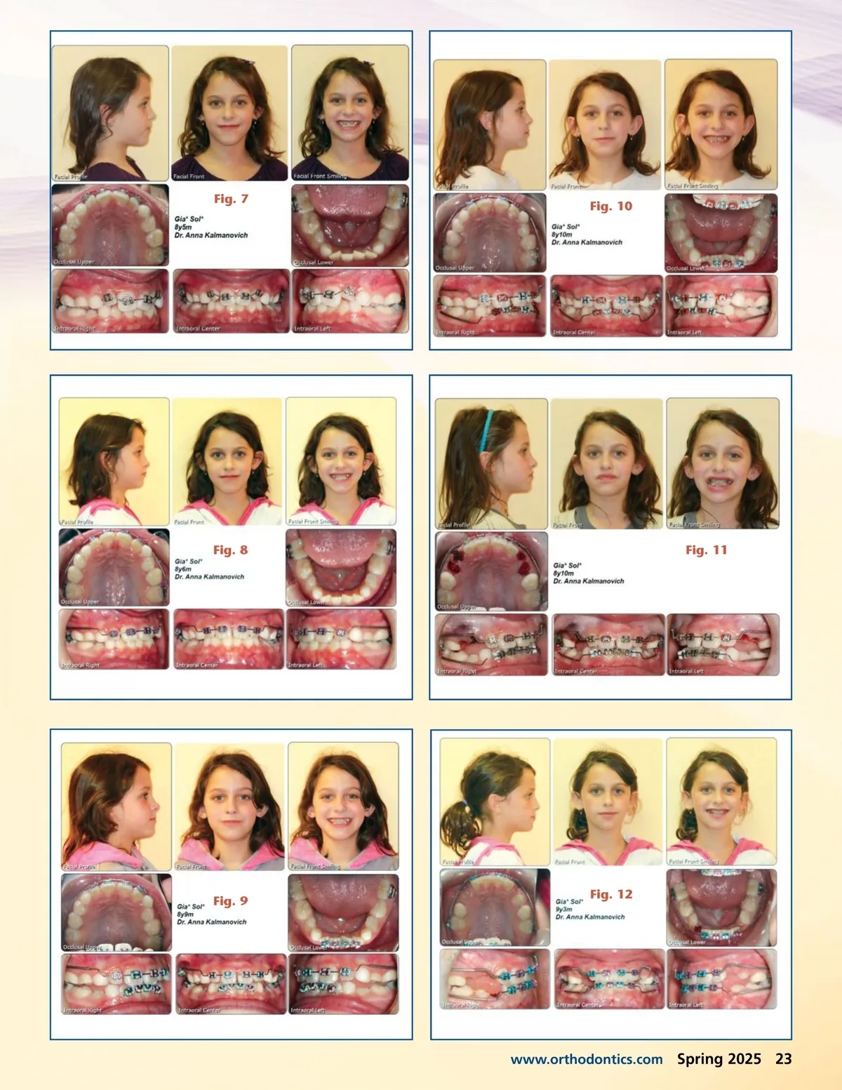
Fig. 5 Fig. 3 Fig. 4b Fig. 6 Fig. 4a Progress Treatment was sectioned into two stages as follows: Stage 1 ᕡ Treatment began when the patient was 7 years old. A rapid palate expanderwas placed for two months (Fig. 3); a facemask was incorporated once the poste-rior crossbite was corrected (Figs. 4a and 4b). Three months later, the patient’s sagittal crossbite was corrected (Fig. 5). ᕢ An accelerated lateral incisor eruption occurred with the extractions of teeth D and G and laser exposure of teeth 7 and 10 (Fig. 6). This allowed us to continue arch development with the utility arch wire, because the timeframe for a successful non-surgical approach is limited. In the case of severe maxillary constriction, we anticipate the delay of eruption of permanent upper cuspids. The best time is when one half to two thirds of the permanent roots are formed. ᕣ The approach of bracketing with an upper sectional wire is easier than other approaches on the patient and causes less pain for transitioning to the utility arch wire (Fig. 7). ᕤ A utility arch was added 1 month after placing the sectional wire. At this point, the teeth were already straighter than before (Fig. 8). ᕥ We placed a lower sectional wire and composite buttons on the lower D teeth (Fig. 9). ᕦ We then added the lower utility arch wire (Fig.10). ᕧ We began serial guidance with extractions of teeth B, C, H, and I to promote favorable eruption of the permanent dentition (Fig. 11). ᕨ We then extracted the lower right C tooth. After the patient lost her lower left C, we extracted the lower right C to maintain her midline (Fig. 12). ᕩ After 24 months at which time the patient was 9 years old, we removed the brackets, took final records for completion of phase 1, and placed retainers (Fig. 13). Stage 2 ᕡ Two years after initial stage completion, we bracketed the 22 Spring 2025 JAOS
Journal of the American Orthodontic Society Spring 2025: Page 22