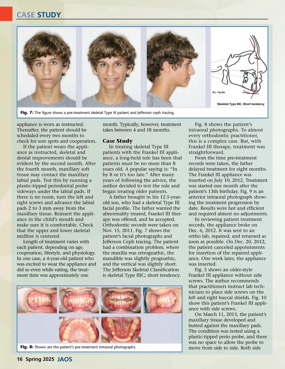
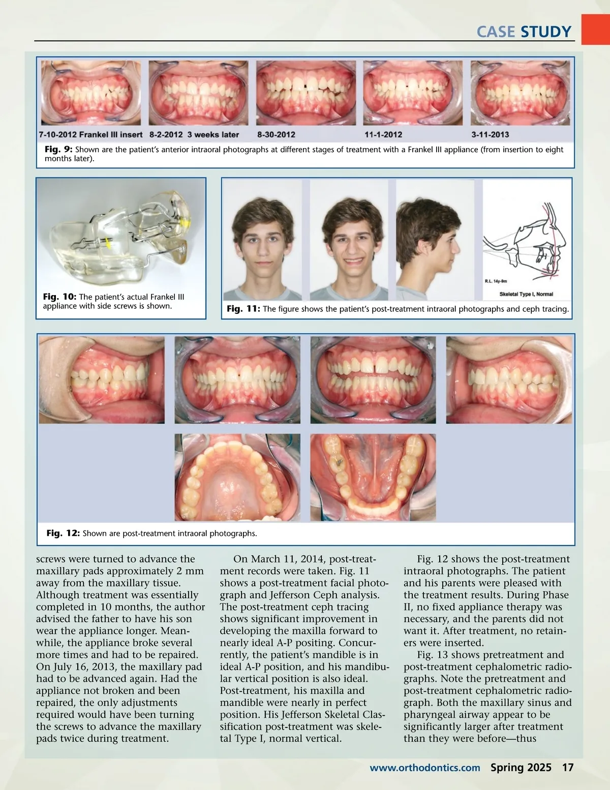
CASE STUDY Fig. 7: The figure shows a pre-treatment skeletal Type III patient and Jefferson ceph tracing. appliance is worn as instructed. Thereafter, the patient should be scheduled every two months to check for sore spots and cooperation. If the patient wears the appli-ance as instructed, skeletal and dental improvements should be evident by the second month. After the fourth month, maxillary soft tissue may contact the maxillary labial pads. Test this by running a plastic-tipped periodontal probe sideways under the labial pads. If there is no room, turn the left and right screws and advance the labial pads 2 to 3 mm away from the maxillary tissue. Reinsert the appli-ance in the child’s mouth and make sure it is comfortable. Check that the upper and lower skeletal midline is centered. Length of treatment varies with each patient, depending on age, cooperation, lifestyle, and physiology. In one case, a 4-year-old patient who was excited to wear the appliance and did so even while eating, the treat-ment time was approximately one month. Typically, however, treatment takes between 4 and 18 months. Case Study In treating skeletal Type III patients with the Frankel III appli-ance, a long-held rule has been that patients must be no more than 8 years old. A popular saying is: “In by 8 or it’s too late.” After many years of following the advice, the author decided to test the rule and began treating older patients. A father brought in his 12.5-year-old son, who had a skeletal Type III facial profile. The father wanted the abnormality treated, Frankel III ther-apy was offered, and he accepted. Orthodontic records were taken on Nov. 15, 2011. Fig. 7 shows the patient’s facial photographs and Jefferson Ceph tracing. The patient had a combination problem, where the maxilla was retrognathic, the mandible was slightly prognathic, and the vertical was slightly short. The Jefferson Skeletal Classification is skeletal Type IIIC; short tendency. Fig. 8: Shown are the patient’s pre-treatment intraoral photographs. Fig. 8 shows the patient’s intraoral photographs. To almost every orthodontic practitioner, this is a complex case. But, with Frankel III therapy, treatment was straightforward. From the time pre-treatment records were taken, the father delayed treatment for eight months. The Frankel III appliance was inserted on July 10, 2012. Treatment was started one month after the patient’s 13th birthday. Fig. 9 is an anterior intraoral photograph show-ing the treatment progression by date. Results were fast and efficient and required almost no adjustments. In reviewing patient treatment records, the appliance broke on Dec. 6, 2012. It was sent to an ortho lab, repaired, and returned as soon as possible. On Dec. 20, 2012, the patient canceled appointments for insertion of the repaired appli-ance. One week later, the appliance was inserted. Fig. 3 shows an older-style Frankel III appliance without side screws. The author recommends that practitioners instruct lab tech-nicians to place side screws on the left and right buccal shields. Fig. 10 show this patient’s Frankel III appli-ance with side screws. On March 11, 2013, the patient’s maxillary tissue developed and butted against the maxillary pads. The condition was tested using a plastic-tipped perio probe, and there was no space to allow the probe to move from side to side. Both side 16 Spring 2025 JAOS
Journal of the American Orthodontic Society Spring 2025: Page 16