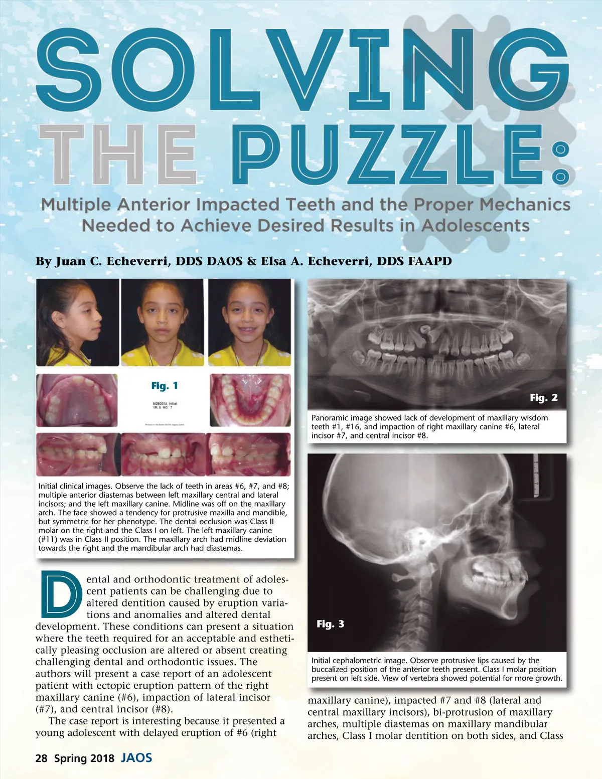
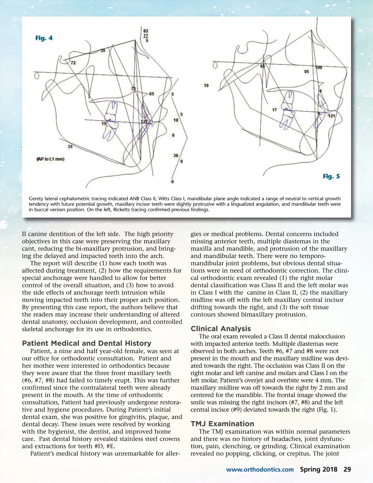
By Juan C. Echeverri, DDS DAOS & Elsa A. Echeverri, DDS FAAPD Fig. 1 Fig. 2 Panoramic image showed lack of development of maxillary wisdom teeth #1, #16, and impaction of right maxillary canine #6, lateral incisor #7, and central incisor #8. Initial clinical images. Observe the lack of teeth in areas #6, #7, and #8; multiple anterior diastemas between left maxillary central and lateral incisors; and the left maxillary canine. Midline was off on the maxillary arch. The face showed a tendency for protrusive maxilla and mandible, but symmetric for her phenotype. The dental occlusion was Class II molar on the right and the Class I on left. The left maxillary canine (#11) was in Class II position. The maxillary arch had midline deviation towards the right and the mandibular arch had diastemas. ental and orthodontic treatment of adoles-cent patients can be challenging due to altered dentition caused by eruption varia-tions and anomalies and altered dental development. These conditions can present a situation where the teeth required for an acceptable and estheti-cally pleasing occlusion are altered or absent creating challenging dental and orthodontic issues. The authors will present a case report of an adolescent patient with ectopic eruption pattern of the right maxillary canine (#6), impaction of lateral incisor (#7), and central incisor (#8). The case report is interesting because it presented a young adolescent with delayed eruption of #6 (right D Fig. 3 Initial cephalometric image. Observe protrusive lips caused by the buccalized position of the anterior teeth present. Class I molar position present on left side. View of vertebra showed potential for more growth. maxillary canine), impacted #7 and #8 (lateral and central maxillary incisors), bi-protrusion of maxillary arches, multiple diastemas on maxillary mandibular arches, Class I molar dentition on both sides, and Class 28 Spring 2018 JAOS
Journal of the American Orthodontic Society Spring 2018: Page 28