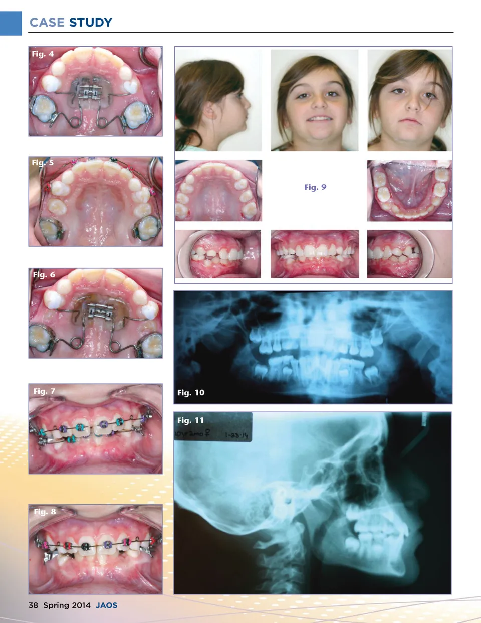
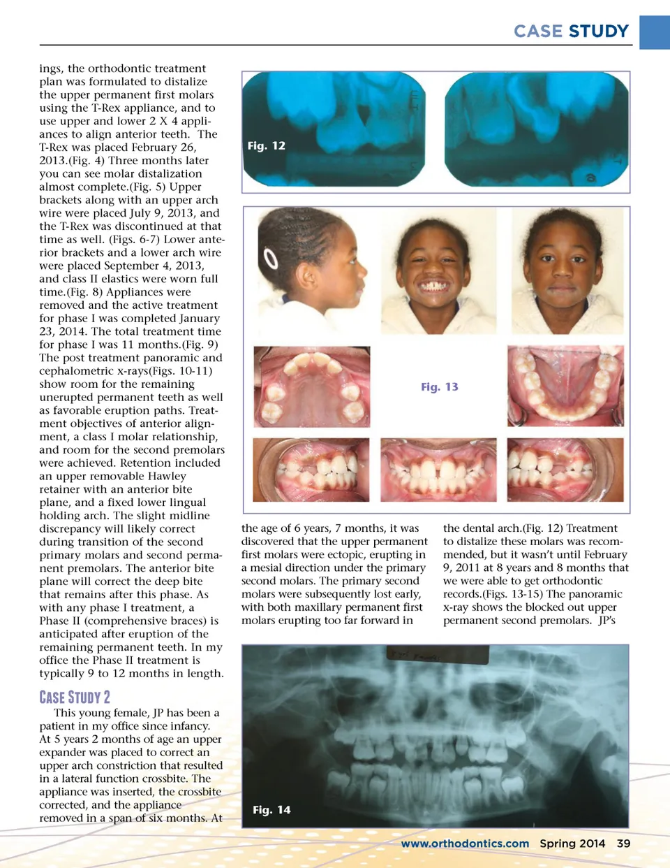
CASE STUDY ings, the orthodontic treatment plan was formulated to distalize the upper permanent first molars using the T-Rex appliance, and to use upper and lower 2 X 4 appli-ances to align anterior teeth. The T-Rex was placed February 26, 2013.(Fig. 4) Three months later you can see molar distalization almost complete.(Fig. 5) Upper brackets along with an upper arch wire were placed July 9, 2013, and the T-Rex was discontinued at that time as well. (Figs. 6-7) Lower ante-rior brackets and a lower arch wire were placed September 4, 2013, and class II elastics were worn full time.(Fig. 8) Appliances were removed and the active treatment for phase I was completed January 23, 2014. The total treatment time for phase I was 11 months.(Fig. 9) The post treatment panoramic and cephalometric x-rays(Figs. 10-11) show room for the remaining unerupted permanent teeth as well as favorable eruption paths. Treat-ment objectives of anterior align-ment, a class I molar relationship, and room for the second premolars were achieved. Retention included an upper removable Hawley retainer with an anterior bite plane, and a fixed lower lingual holding arch. The slight midline discrepancy will likely correct during transition of the second primary molars and second perma-nent premolars. The anterior bite plane will correct the deep bite that remains after this phase. As with any phase I treatment, a Phase II (comprehensive braces) is anticipated after eruption of the remaining permanent teeth. In my office the Phase II treatment is typically 9 to 12 months in length. Fig. 12 Fig. 13 the age of 6 years, 7 months, it was discovered that the upper permanent first molars were ectopic, erupting in a mesial direction under the primary second molars. The primary second molars were subsequently lost early, with both maxillary permanent first molars erupting too far forward in the dental arch.(Fig. 12) Treatment to distalize these molars was recom-mended, but it wasn’t until February 9, 2011 at 8 years and 8 months that we were able to get orthodontic records.(Figs. 13-15) The panoramic x-ray shows the blocked out upper permanent second premolars. JP’s �d;d;a; !�b;f;�e;�f;! This young female, JP has been a patient in my office since infancy. At 5 years 2 months of age an upper expander was placed to correct an upper arch constriction that resulted in a lateral function crossbite. The appliance was inserted, the crossbite corrected, and the appliance removed in a span of six months. At Fig. 14 www.orthodontics.com Spring 2014 39
Journal of the American Orthodontic Society Spring 2014: Page 39