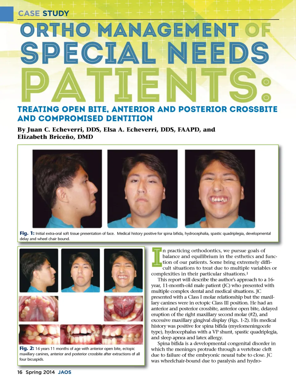
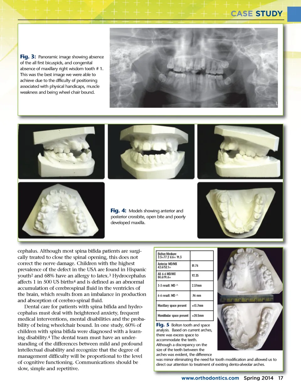
CASE STUDY Treating Open Bite, Anterior and Posterior Crossbite and Compromised Dentition By Juan C. Echeverri, DDS, Elsa A. Echeverri, DDS, FAAPD, and Elizabeth Briceño, DMD Fig. 1: Initial extra-oral soft tissue presentation of face. Medical history positive for spina bifida, hydrocephalia, spastic quadriplegia, developmental delay and wheel chair bound. I Fig. 2: 16 years 11 months of age with anterior open bite, ectopic maxillary canines, anterior and posterior crossbite after extractions of all four bicuspids. 16 Spring 2014 J JAOS AOS n practicing orthodontics, we pursue goals of balance and equilibrium in the esthetics and func-tion of our patients. Some bring extremely diffi-cult situations to treat due to multiple variables or complexities in their particular situations. 1 This report will describe the author’s approach to a 16-year, 11-month-old male patient (JC) who presented with multiple complex dental and medical situations. JC presented with a Class I molar relationship but the maxil-lary canines were in ectopic Class III position. He had an anterior and posterior crossbite, anterior open bite, delayed eruption of the right maxillary second molar (#2), and excessive maxillary gingival display (Figs. 1-2). His medical history was positive for spina bifida (myelomeningocele type), hydrocephalus with a VP shunt, spastic quadriplegia, and sleep apnea and latex allergy. Spina bifida is a developmental congenital disorder in which the meninges protrude through a vertebrae cleft due to failure of the embryonic neural tube to close. JC was wheelchair-bound due to paralysis and hydro-
Journal of the American Orthodontic Society Spring 2014: Page 16