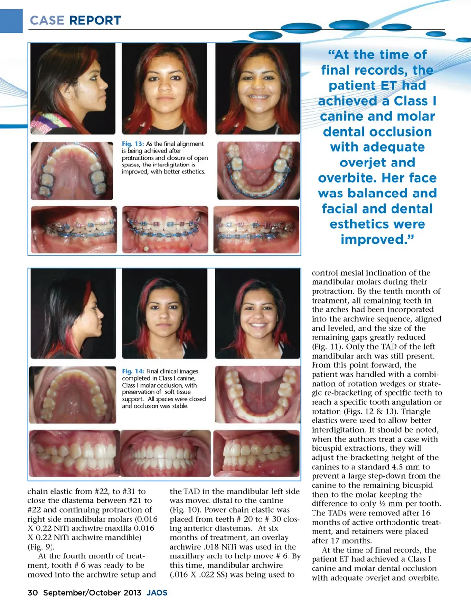
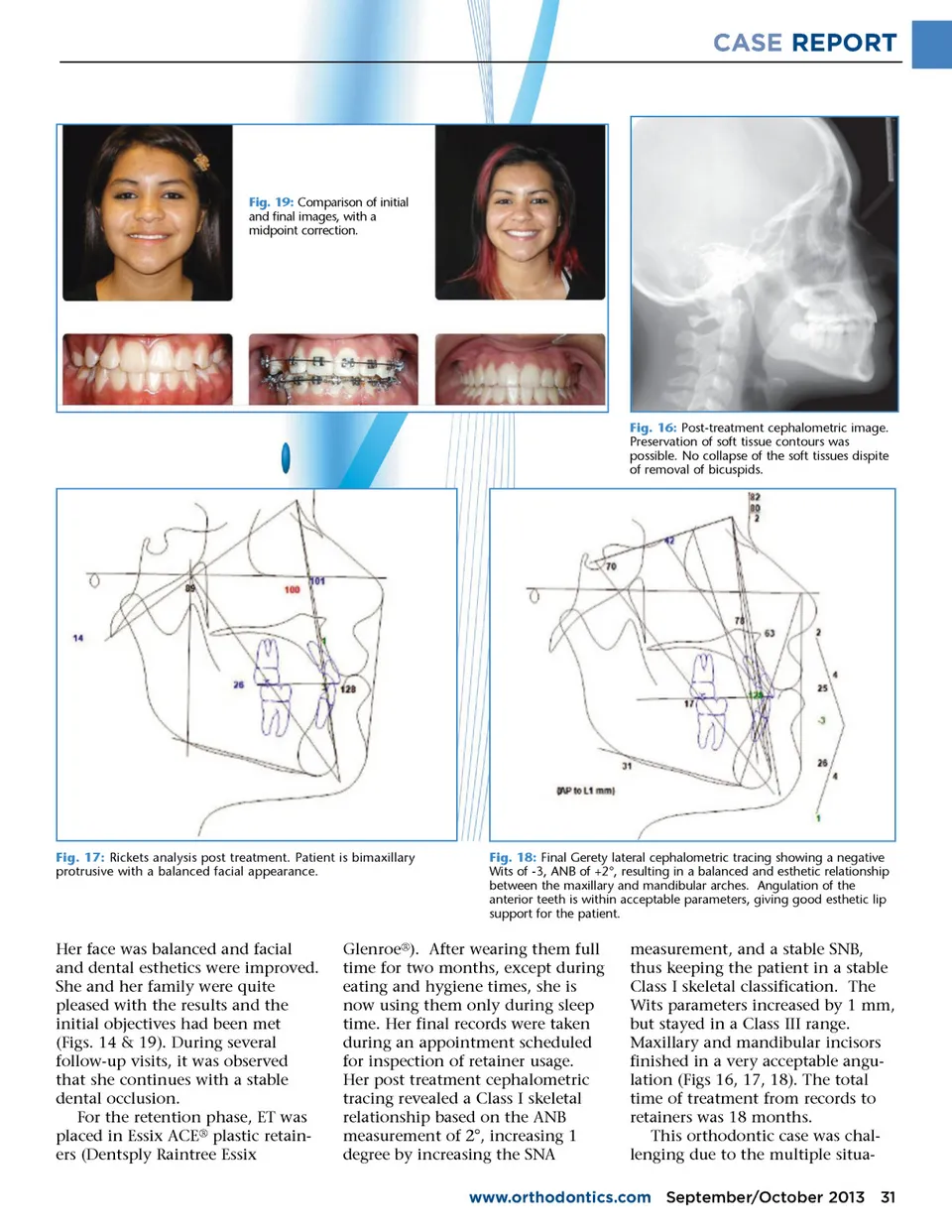
CASE REPORT Fig. 19: Comparison of initial and final images, with a midpoint correction. Fig. 16: Post-treatment cephalometric image. Preservation of soft tissue contours was possible. No collapse of the soft tissues dispite of removal of bicuspids. Fig. 17: Rickets analysis post treatment. Patient is bimaxillary protrusive with a balanced facial appearance. Fig. 18: Final Gerety lateral cephalometric tracing showing a negative Wits of -3, ANB of +2°, resulting in a balanced and esthetic relationship between the maxillary and mandibular arches. Angulation of the anterior teeth is within acceptable parameters, giving good esthetic lip support for the patient. Her face was balanced and facial and dental esthetics were improved. She and her family were quite pleased with the results and the initial objectives had been met (Figs. 14 & 19). During several follow-up visits, it was observed that she continues with a stable dental occlusion. For the retention phase, ET was placed in Essix ACE ® plastic retain-ers (Dentsply Raintree Essix Glenroe ® ). After wearing them full time for two months, except during eating and hygiene times, she is now using them only during sleep time. Her final records were taken during an appointment scheduled for inspection of retainer usage. Her post treatment cephalometric tracing revealed a Class I skeletal relationship based on the ANB measurement of 2°, increasing 1 degree by increasing the SNA measurement, and a stable SNB, thus keeping the patient in a stable Class I skeletal classification. The Wits parameters increased by 1 mm, but stayed in a Class III range. Maxillary and mandibular incisors finished in a very acceptable angu-lation (Figs 16, 17, 18). The total time of treatment from records to retainers was 18 months. This orthodontic case was chal-lenging due to the multiple situa-www.orthodontics.com September/October 2013 31
Journal of the American Orthodontic Society September-October 2013: Page 31