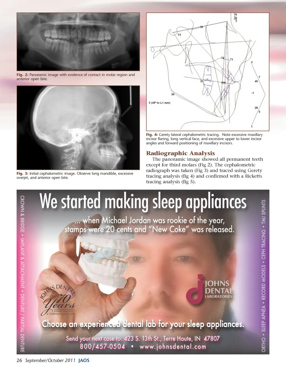
Fig. 2: Panoramic image with evidence of contact in molar region and anterior open bite. Fig. 4: Gerety lateral cephalometric tracing. Note excessive maxillary incisor flaring, long vertical face, and excessive upper to lower incisor angles and forward positioning of maxillary incisors. Radiographic Analysis The panoramic image showed all permanent teeth except for third molars (Fig 2). The cephalometric radiograph was taken (Fig 3) and traced using Gerety tracing analysis (fig 4) and confirmed with a Ricketts tracing analysis (fig 5). Fig. 3: Initial cephalometric image. Observe long mandible, excessive overjet, and anterior open bite. 26 September/October 2011 JAOS
Journal of the American Orthodontic Society September-October 2011: Page 26