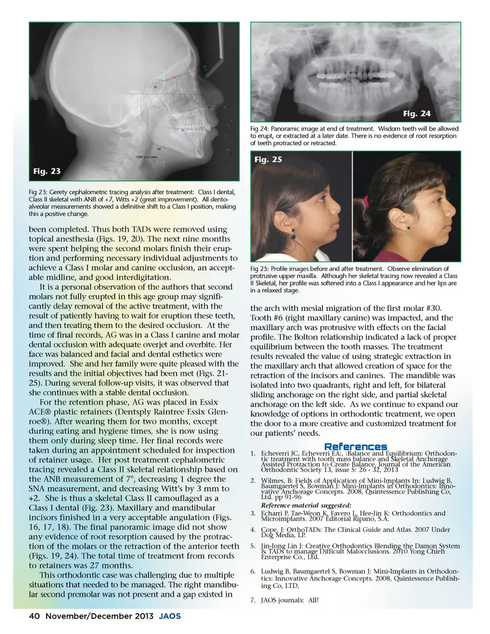
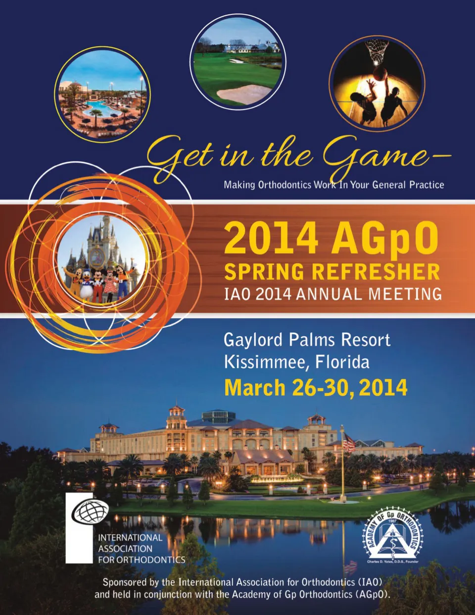
Fig. 24 Fig 24: Panoramic image at end of treatment. Wisdom teeth will be allowed to erupt, or extracted at a later date. There is no evidence of root resorption of teeth protracted or retracted. Fig. 25 Fig. 23 Fig 23: Gerety cephalometric tracing analysis after treatment: Class I dental, Class II skeletal with ANB of +7, Witts +2 (great improvement). All dento-alveolar measurements showed a definitive shift to a Class I position, making this a positive change. been completed. Thus both TADs were removed using topical anesthesia (Figs. 19, 20). The next nine months were spent helping the second molars finish their erup-tion and performing necessary individual adjustments to achieve a Class I molar and canine occlusion, an accept-able midline, and good interdigitation. It is a personal observation of the authors that second molars not fully erupted in this age group may signifi-cantly delay removal of the active treatment, with the result of patiently having to wait for eruption these teeth, and then treating them to the desired occlusion. At the time of final records, AG was in a Class I canine and molar dental occlusion with adequate overjet and overbite. Her face was balanced and facial and dental esthetics were improved. She and her family were quite pleased with the results and the initial objectives had been met (Figs. 21-25). During several follow-up visits, it was observed that she continues with a stable dental occlusion. For the retention phase, AG was placed in Essix ACE® plastic retainers (Dentsply Raintree Essix Glen-roe®). After wearing them for two months, except during eating and hygiene times, she is now using them only during sleep time. Her final records were taken during an appointment scheduled for inspection of retainer usage. Her post treatment cephalometric tracing revealed a Class II skeletal relationship based on the ANB measurement of 7°, decreasing 1 degree the SNA measurement, and decreasing Witt’s by 3 mm to +2. She is thus a skeletal Class II camouflaged as a Class I dental (Fig. 23). Maxillary and mandibular incisors finished in a very acceptable angulation (Figs. 16, 17, 18). The final panoramic image did not show any evidence of root resorption caused by the protrac-tion of the molars or the retraction of the anterior teeth (Figs. 19, 24). The total time of treatment from records to retainers was 27 months. This orthodontic case was challenging due to multiple situations that needed to be managed. The right mandibu-lar second premolar was not present and a gap existed in 40 November/December 2013 JAOS Fig 25: Profile images before and after treatment. Observe elimination of protrusive upper maxilla. Although her skeletal tracing now revealed a Class II Skeletal, her profile was softened into a Class I appearance and her lips are in a relaxed stage. the arch with mesial migration of the first molar #30. Tooth #6 (right maxillary canine) was impacted, and the maxillary arch was protrusive with effects on the facial profile. The Bolton relationship indicated a lack of proper equilibrium between the tooth masses. The treatment results revealed the value of using strategic extraction in the maxillary arch that allowed creation of space for the retraction of the incisors and canines. The mandible was isolated into two quadrants, right and left, for bilateral sliding anchorage on the right side, and partial skeletal anchorage on the left side. As we continue to expand our knowledge of options in orthodontic treatment, we open the door to a more creative and customized treatment for our patients’ needs. 1. Echeverri JC, Echeverri EA:, :Balance and Equilibrium: Orthodon-tic treatment with tooth mass balance and Skeletal Anchorage Assisted Protraction to Create Balance, Journal of the American Orthodontic Society 13, issue 5: 26 -32, 2013 2. Wilmes, B: Fields of Application of Mini-Implants In: Ludwig B, Baumgaertel S, Bowman J: Mini-Implants in Orthodontics: Inno-vative Anchorage Concepts. 2008, Quintessence Publishing Co, Ltd. pp 91-96 Reference material suggested: 3. Echarri P, Tae-Weon K, Favero L, Hee-Jin K: Orthodontics and Microimplants. 2007 Editorial Ripano, S.A. 4. Cope, J: OrthoTADs: The Clinical Guide and Atlas. 2007 Under Dog Media, LP. 5. Jin-Jong Lin J: Creative Orthodontics Blending the Damon System & TADs to manage Difficult Malocclusions. 2010 Yong Chieh Enterprise Co., Ltd. 6. Ludwig B, Baumgaertel S, Bowman J: Mini-Implants in Orthodon-tics: Innovative Anchorage Concepts. 2008, Quintessence Publish-ing Co, LTD, 7. JAOS journals: All! ! !d;!"!e;
Journal of the American Orthodontic Society November-December 2013: Page 40