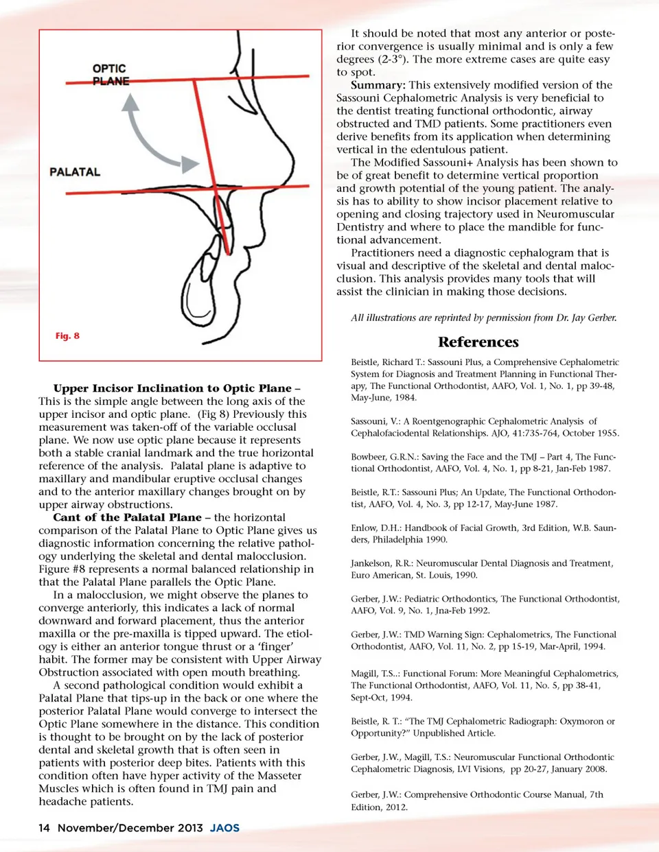
It should be noted that most any anterior or poste-rior convergence is usually minimal and is only a few degrees (2-3°). The more extreme cases are quite easy to spot. Summary: This extensively modified version of the Sassouni Cephalometric Analysis is very beneficial to the dentist treating functional orthodontic, airway obstructed and TMD patients. Some practitioners even derive benefits from its application when determining vertical in the edentulous patient. The Modified Sassouni+ Analysis has been shown to be of great benefit to determine vertical proportion and growth potential of the young patient. The analy-sis has to ability to show incisor placement relative to opening and closing trajectory used in Neuromuscular Dentistry and where to place the mandible for func-tional advancement. Practitioners need a diagnostic cephalogram that is visual and descriptive of the skeletal and dental maloc-clusion. This analysis provides many tools that will assist the clinician in making those decisions. All illustrations are reprinted by permission from Dr. Jay Gerber. Fig. 8 References Beistle, Richard T.: Sassouni Plus, a Comprehensive Cephalometric System for Diagnosis and Treatment Planning in Functional Ther-apy, The Functional Orthodontist, AAFO, Vol. 1, No. 1, pp 39-48, May-June, 1984. Sassouni, V.: A Roentgenographic Cephalometric Analysis of Cephalofaciodental Relationships. AJO, 41:735-764, October 1955. Bowbeer, G.R.N.: Saving the Face and the TMJ – Part 4, The Func-tional Orthodontist, AAFO, Vol. 4, No. 1, pp 8-21, Jan-Feb 1987. Beistle, R.T.: Sassouni Plus; An Update, The Functional Orthodon-tist, AAFO, Vol. 4, No. 3, pp 12-17, May-June 1987. Enlow, D.H.: Handbook of Facial Growth, 3rd Edition, W.B. Saun-ders, Philadelphia 1990. Jankelson, R.R.: Neuromuscular Dental Diagnosis and Treatment, Euro American, St. Louis, 1990. Gerber, J.W.: Pediatric Orthodontics, The Functional Orthodontist, AAFO, Vol. 9, No. 1, Jna-Feb 1992. Gerber, J.W.: TMD Warning Sign: Cephalometrics, The Functional Orthodontist, AAFO, Vol. 11, No. 2, pp 15-19, Mar-April, 1994. Magill, T.S..: Functional Forum: More Meaningful Cephalometrics, The Functional Orthodontist, AAFO, Vol. 11, No. 5, pp 38-41, Sept-Oct, 1994. Beistle, R. T.: “The TMJ Cephalometric Radiograph: Oxymoron or Opportunity?” Unpublished Article. Gerber, J.W., Magill, T.S.: Neuromuscular Functional Orthodontic Cephalometric Diagnosis, LVI Visions, pp 20-27, January 2008. Gerber, J.W.: Comprehensive Orthodontic Course Manual, 7th Edition, 2012. Upper Incisor Inclination to Optic Plane – This is the simple angle between the long axis of the upper incisor and optic plane. (Fig 8) Previously this measurement was taken-off of the variable occlusal plane. We now use optic plane because it represents both a stable cranial landmark and the true horizontal reference of the analysis. Palatal plane is adaptive to maxillary and mandibular eruptive occlusal changes and to the anterior maxillary changes brought on by upper airway obstructions. Cant of the Palatal Plane – the horizontal comparison of the Palatal Plane to Optic Plane gives us diagnostic information concerning the relative pathol-ogy underlying the skeletal and dental malocclusion. Figure #8 represents a normal balanced relationship in that the Palatal Plane parallels the Optic Plane. In a malocclusion, we might observe the planes to converge anteriorly, this indicates a lack of normal downward and forward placement, thus the anterior maxilla or the pre-maxilla is tipped upward. The etiol-ogy is either an anterior tongue thrust or a ‘finger’ habit. The former may be consistent with Upper Airway Obstruction associated with open mouth breathing. A second pathological condition would exhibit a Palatal Plane that tips-up in the back or one where the posterior Palatal Plane would converge to intersect the Optic Plane somewhere in the distance. This condition is thought to be brought on by the lack of posterior dental and skeletal growth that is often seen in patients with posterior deep bites. Patients with this condition often have hyper activity of the Masseter Muscles which is often found in TMJ pain and headache patients. 14 November/December 2013 JAOS
Journal of the American Orthodontic Society November-December 2013: Page 14