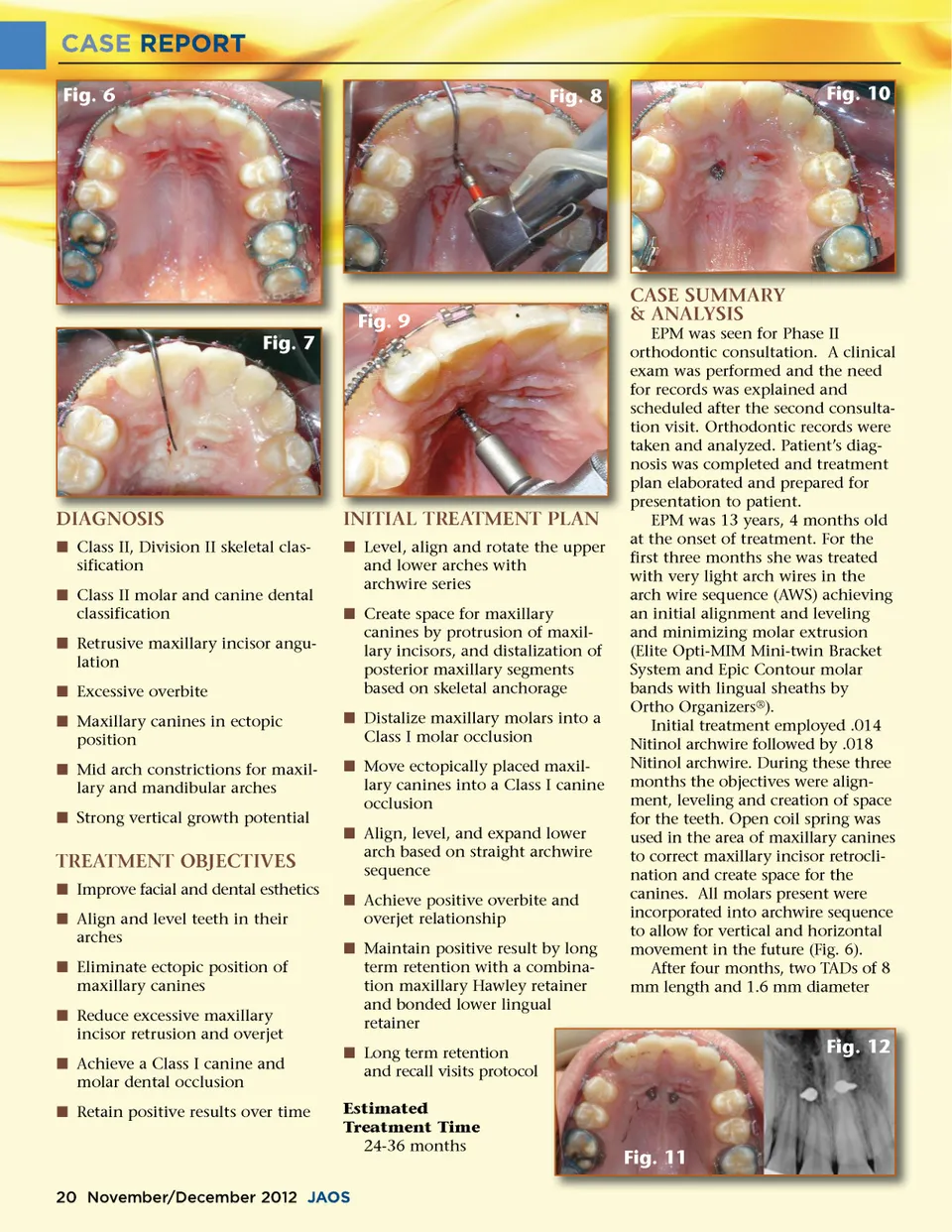
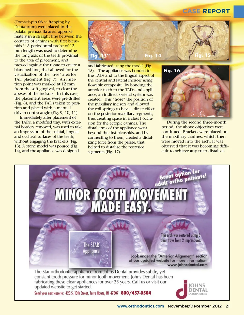
CASE REPORT (Tomas ® -pin 08 selftapping by Dentaurum) were placed in the palatal premaxilla area, approxi-mately in a straight line between the contacts of canines with first bicus-pids. 11 A periodontal probe of 12 mm length was used to determine the long axis of the teeth proximal to the area of placement, and pressed against the tissue to create a blanched line, that allowed for the visualization of the “free” area for TAD placement (Fig. 7). An inser-tion point was marked at 12 mm from the soft gingival, to clear the apexes of the incisors. In this case, the placement areas were pre-drilled (Fig. 8), and the TADs taken to posi-tion and placed with a manual driven contra-angle (Fig. 9, 10, 11). Immediately after placement of the TADs, a modified tray, with exter-nal borders removed, was used to take an impression of the palatal, lingual and occlusal surfaces of the teeth, without engaging the brackets (Fig. 13). A stone model was poured (Fig. 14), and the appliance was designed Fig. 13 Fig. 14 Fig. 16 Fig. 15 fabricated and fa f bricated using the model (Fig. (Fig 15). The appliance was bonded to the TADs and to the lingual aspect of the central and lateral incisors using flowable composite. By bonding the anterior teeth to the TADs and appli-ance, an indirect skeletal system was created. This “froze” the position of the maxillary incisors and allowed the coil springs to have a direct effect on the posterior maxillary segments, thus creating space in a class I occlu-sion for the ectopic canines. The distal arms of the appliance went beyond the first bicuspids, and by connecting to them, created a distal-izing force from the palate, that helped to distalize the posterior segments (Fig. 17). During the second three month three-month period, the above objectives were continued. Brackets were placed on the maxillary canines, which then were moved into the arch. It was observed that it was becoming diffi-cult to achieve any truer distaliza-www.orthodontics.com November/December 2012 21
Journal of the American Orthodontic Society November-December 2012: Page 21