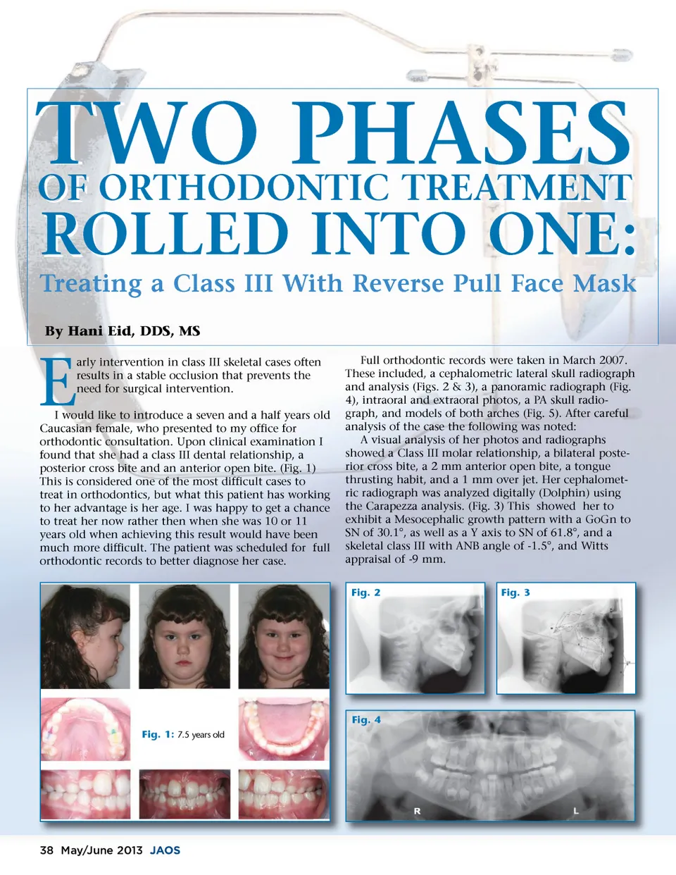
TWO PHASES OF ORTHODONTIC TREATMENT By Hani Eid, DDS, MS ROLLED INTO ONE: arly intervention in class III skeletal cases often results in a stable occlusion that prevents the need for surgical intervention. Full orthodontic records were taken in March 2007. These included, a cephalometric lateral skull radiograph and analysis (Figs. 2 & 3), a panoramic radiograph (Fig. 4), intraoral and extraoral photos, a PA skull radio-graph, and models of both arches (Fig. 5). After careful analysis of the case the following was noted: A visual analysis of her photos and radiographs showed a Class III molar relationship, a bilateral poste-rior cross bite, a 2 mm anterior open bite, a tongue thrusting habit, and a 1 mm over jet. Her cephalomet-ric radiograph was analyzed digitally (Dolphin) using the Carapezza analysis. (Fig. 3) This showed her to exhibit a Mesocephalic growth pattern with a GoGn to SN of 30.1°, as well as a Y axis to SN of 61.8°, and a skeletal class III with ANB angle of -1.5°, and Witts appraisal of -9 mm. Fig. 2 Fig. 3 Treating a Class III With Reverse Pull Face Mask E I would like to introduce a seven and a half years old Caucasian female, who presented to my office for orthodontic consultation. Upon clinical examination I found that she had a class III dental relationship, a posterior cross bite and an anterior open bite. (Fig. 1) This is considered one of the most difficult cases to treat in orthodontics, but what this patient has working to her advantage is her age. I was happy to get a chance to treat her now rather then when she was 10 or 11 years old when achieving this result would have been much more difficult. The patient was scheduled for full orthodontic records to better diagnose her case. Fig. 4 Fig. 1: 7.5 years old 38 May/June 2013 JAOS
Journal of the American Orthodontic Society May-June 2013: Page 38