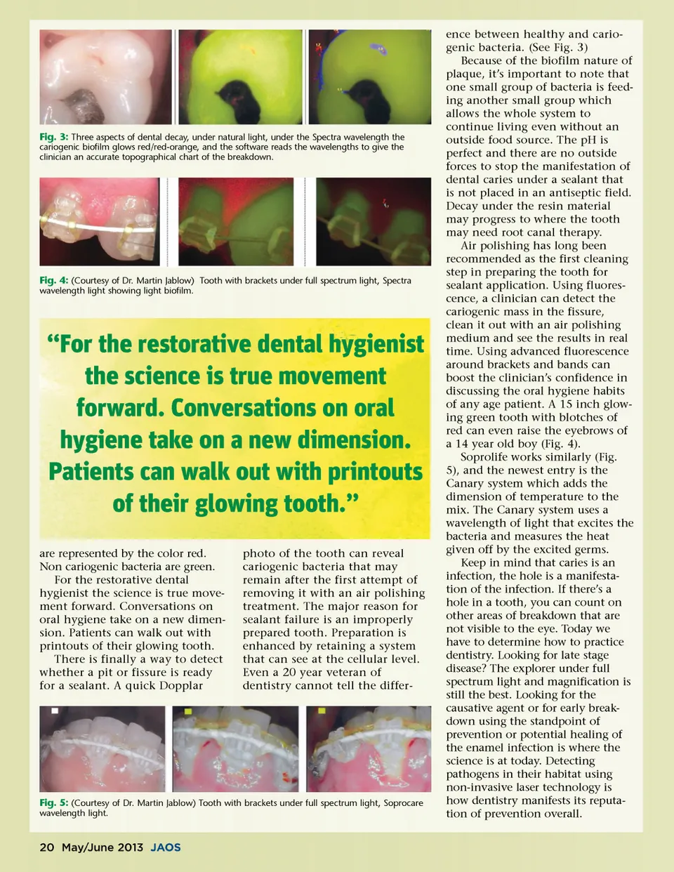
Fig. 3: Three aspects of dental decay, under natural light, under the Spectra wavelength the cariogenic biofilm glows red/red-orange, and the software reads the wavelengths to give the clinician an accurate topographical chart of the breakdown. Fig. 4: (Courtesy of Dr. Martin Jablow) Tooth with brackets under full spectrum light, Spectra wavelength light showing light biofilm. “For the restorative dental hygienist the science is true movement forward. Conversations on oral hygiene take on a new dimension. Patients can walk out with printouts of their glowing tooth.” are represented by the color red. Non cariogenic bacteria are green. For the restorative dental hygienist the science is true move-ment forward. Conversations on oral hygiene take on a new dimen-sion. Patients can walk out with printouts of their glowing tooth. There is finally a way to detect whether a pit or fissure is ready for a sealant. A quick Dopplar photo of the tooth can reveal cariogenic bacteria that may remain after the first attempt of removing it with an air polishing treatment. The major reason for sealant failure is an improperly prepared tooth. Preparation is enhanced by retaining a system that can see at the cellular level. Even a 20 year veteran of dentistry cannot tell the differ-Fig. 5: (Courtesy of Dr. Martin Jablow) Tooth with brackets under full spectrum light, Soprocare wavelength light. ence between healthy and cario-genic bacteria. (See Fig. 3) Because of the biofilm nature of plaque, it’s important to note that one small group of bacteria is feed-ing another small group which allows the whole system to continue living even without an outside food source. The pH is perfect and there are no outside forces to stop the manifestation of dental caries under a sealant that is not placed in an antiseptic field. Decay under the resin material may progress to where the tooth may need root canal therapy. Air polishing has long been recommended as the first cleaning step in preparing the tooth for sealant application. Using fluores-cence, a clinician can detect the cariogenic mass in the fissure, clean it out with an air polishing medium and see the results in real time. Using advanced fluorescence around brackets and bands can boost the clinician’s confidence in discussing the oral hygiene habits of any age patient. A 15 inch glow-ing green tooth with blotches of red can even raise the eyebrows of a 14 year old boy (Fig. 4). Soprolife works similarly (Fig. 5), and the newest entry is the Canary system which adds the dimension of temperature to the mix. The Canary system uses a wavelength of light that excites the bacteria and measures the heat given off by the excited germs. Keep in mind that caries is an infection, the hole is a manifesta-tion of the infection. If there’s a hole in a tooth, you can count on other areas of breakdown that are not visible to the eye. Today we have to determine how to practice dentistry. Looking for late stage disease? The explorer under full spectrum light and magnification is still the best. Looking for the causative agent or for early break-down using the standpoint of prevention or potential healing of the enamel infection is where the science is at today. Detecting pathogens in their habitat using non-invasive laser technology is how dentistry manifests its reputa-tion of prevention overall. 20 May/June 2013 JAOS
Journal of the American Orthodontic Society May-June 2013: Page 20