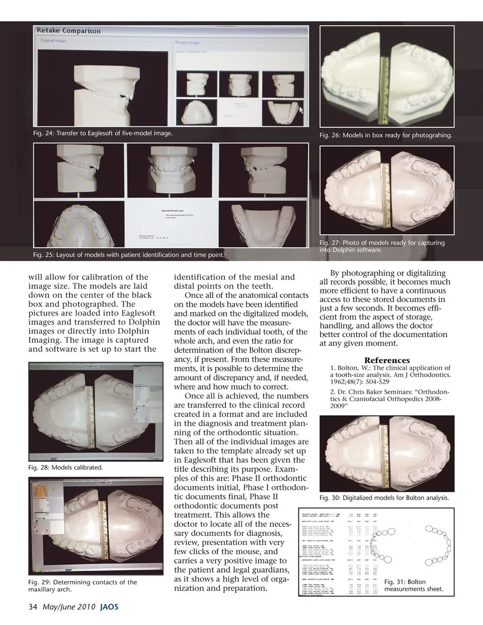
Fig. 24: Transfer to Eaglesoft of five-model image. Fig. 26: Models in box ready for photograhing. Fig. 25: Layout of models with patient identification and time point. will allow for calibration of the image size. The models are laid down on the center of the black box and photographed. The pictures are loaded into Eaglesoft images and transferred to Dolphin images or directly into Dolphin Imaging. The image is captured and software is set up to start the Fig. 28: Models calibrated. Fig. 29: Determining contacts of the maxillary arch. 34 May/June 2010 JAOS identification of the mesial and distal points on the teeth. Once all of the anatomical contacts on the models have been identified and marked on the digitalized models, the doctor will have the measure- ments of each individual tooth, of the whole arch, and even the ratio for determination of the Bolton discrep- ancy, if present. From these measure- ments, it is possible to determine the amount of discrepancy and, if needed, where and how much to correct. Once all is achieved, the numbers are transferred to the clinical record created in a format and are included in the diagnosis and treatment plan- ning of the orthodontic situation. Then all of the individual images are taken to the template already set up in Eaglesoft that has been given the title describing its purpose. Exam- ples of this are: Phase II orthodontic documents initial, Phase I orthodon- tic documents final, Phase II orthodontic documents post treatment. This allows the doctor to locate all of the neces- sary documents for diagnosis, review, presentation with very few clicks of the mouse, and carries a very positive image to the patient and legal guardians, as it shows a high level of orga- nization and preparation. Fig. 27: Photo of models ready for capturing into Dolphin software. By photographing or digitalizing all records possible, it becomes much more efficient to have a continuous access to these stored documents in just a few seconds. It becomes effi- cient from the aspect of storage, handling, and allows the doctor better control of the documentation at any given moment. References 1. Bolton, W.: The clinical application of a tooth-size analysis. Am J Orthodontics. 1962;48(7): 504-529 2. Dr. Chris Baker Seminars: “Orthodon- tics & Craniofacial Orthopedics 2008- 2009” Fig. 30: Digitalized models for Bolton analysis. Fig. 31: Bolton measurements sheet.
Journal of the American Orthodontic Society May-June 2010: Page 34