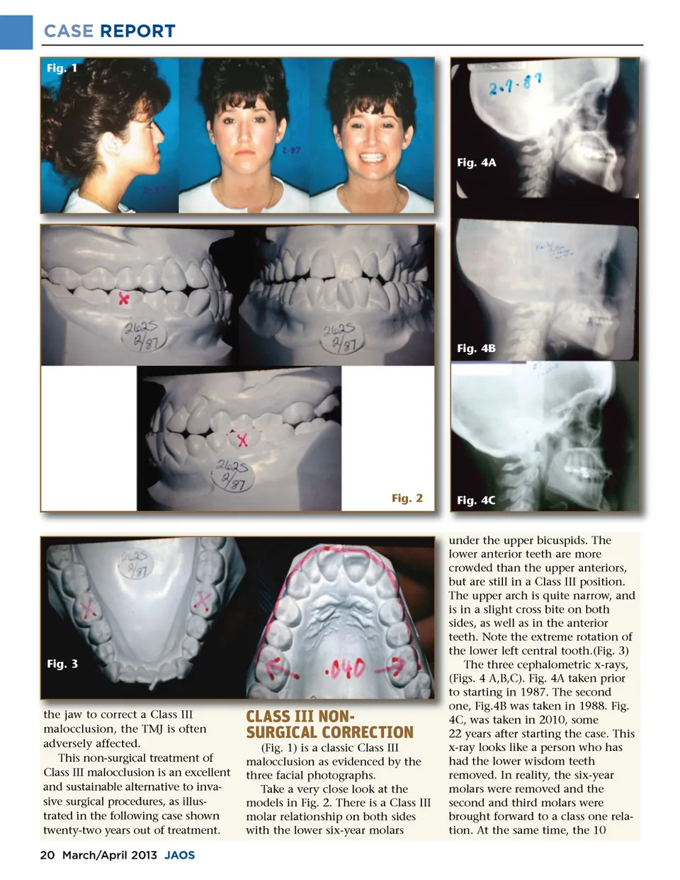
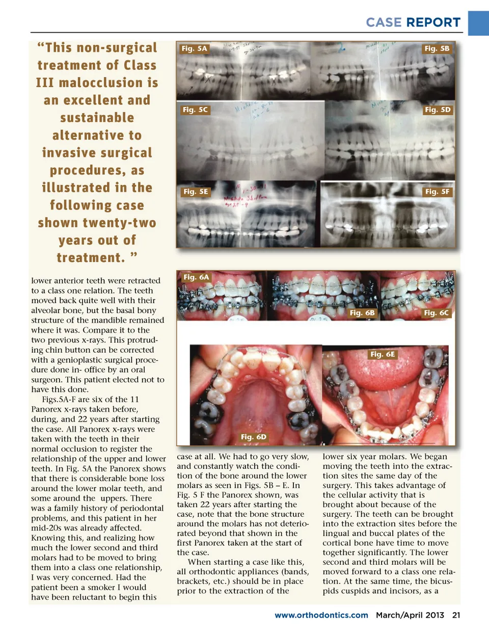
CASE REPORT “This non-surgical treatment of Class III malocclusion is an excellent and sustainable alternative to invasive surgical procedures, as illustrated in the following case shown twenty-two years out of treatment. ” lower anterior teeth were retracted to a class one relation. The teeth moved back quite well with their alveolar bone, but the basal bony structure of the mandible remained where it was. Compare it to the two previous x-rays. This protrud-ing chin button can be corrected with a genioplastic surgical proce-dure done in-office by an oral surgeon. This patient elected not to have this done. Figs.5A-F are six of the 11 Panorex x-rays taken before, during, and 22 years after starting the case. All Panorex x-rays were taken with the teeth in their normal occlusion to register the relationship of the upper and lower teeth. In Fig. 5A the Panorex shows that there is considerable bone loss around the lower molar teeth, and some around the uppers. There was a family history of periodontal problems, and this patient in her mid-20s was already affected. Knowing this, and realizing how much the lower second and third molars had to be moved to bring them into a class one relationship, I was very concerned. Had the patient been a smoker I would have been reluctant to begin this Fig. 5A Fig. 5B Fig. 5C Fig. 5D Fig. 5E Fig. 5F Fig. 6A Fig. 6B Fig. 6C Fig. 6E Fig. 6D W had to go very slow case at all all. We slow, and constantly watch the condi-tion of the bone around the lower molars as seen in Figs. 5B – E. In Fig. 5 F the Panorex shown, was taken 22 years after starting the case, note that the bone structure around the molars has not deterio-rated beyond that shown in the first Panorex taken at the start of the case. When starting a case like this, all orthodontic appliances (bands, brackets, etc.) should be in place prior to the extraction of the molars We W began lower six year molars. moving the teeth into the extrac-tion sites the same day of the surgery. This takes advantage of the cellular activity that is brought about because of the surgery. The teeth can be brought into the extraction sites before the lingual and buccal plates of the cortical bone have time to move together significantly. The lower second and third molars will be moved forward to a class one rela-tion. At the same time, the bicus-pids cuspids and incisors, as a www.orthodontics.com March/April 2013 21
Journal of the American Orthodontic Society March-April 2013: Page 21